介绍
一般状态恶化,发烧,贫血,炎症标志物和肝功能检查升高。
患者资料
年龄:85岁
性别:女
CT
原发性脾淋巴瘤 - 弥漫性大 B 细胞淋巴瘤
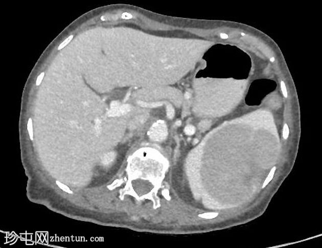
Axial C+ portal venous phase
原发性脾淋巴瘤 - 弥漫性大 B 细胞淋巴瘤
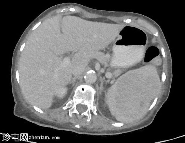
Axial C+ delayed
原发性脾淋巴瘤 - 弥漫性大 B 细胞淋巴瘤
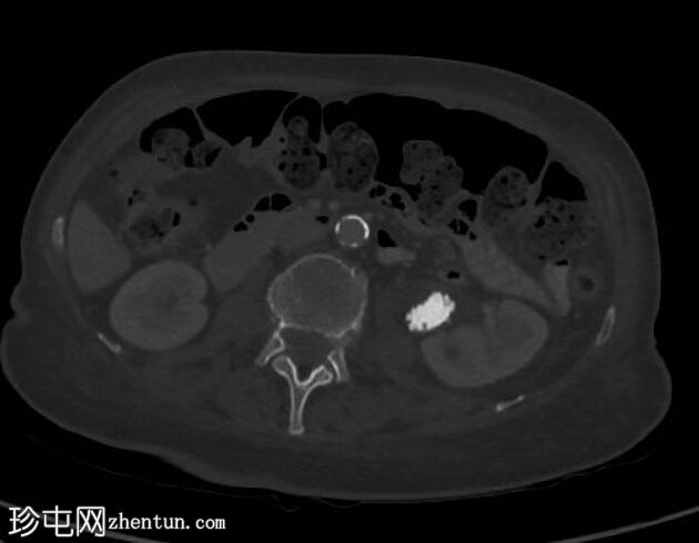
Axial bone window
原发性脾淋巴瘤 - 弥漫性大 B 细胞淋巴瘤
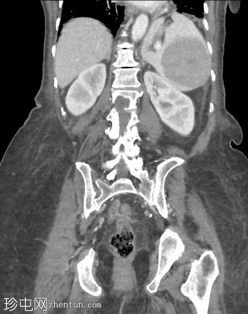
Coronal C+ portal venous phase
原发性脾淋巴瘤 - 弥漫性大 B 细胞淋巴瘤
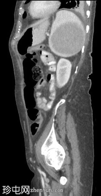
Sagittal C+ portal venous phase
与近 2 年前完成的腹部 CT 相比(见下文研究):
左侧少量胸腔积液。
胆囊切除术和子宫切除术后的状态。
大小为 9.0 x 7.4 x 7.7 cm 的大型低密度低增强脾脏肿块已经显著增大。
胰腺体中的囊性病变,尺寸为 11 x 9 毫米 - 在之前的研究中不可见。
左侧肾外骨盆中的大 Jackstone 结石 - 无变化。
左肾盆旁囊肿。
骨盆内有少量游离腹腔积液。
CT
原发性脾淋巴瘤 - 弥漫性大 B 细胞淋巴瘤
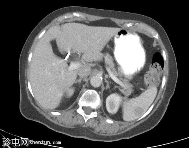
Axial C+ portal venous phase
原发性脾淋巴瘤 - 弥漫性大 B 细胞淋巴瘤
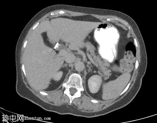
Axial C+ delayed
近 2 年前的腹部 CT:
脾肿块明显较小。
左侧输尿管近端的结石大小相同。
超声波
原发性脾淋巴瘤 - 弥漫性大 B 细胞淋巴瘤
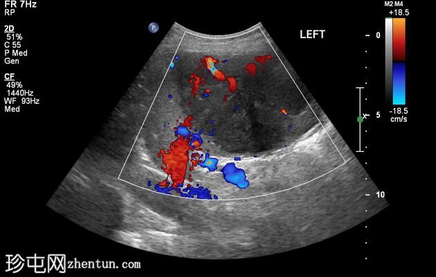
原发性脾淋巴瘤 - 弥漫性大 B 细胞淋巴瘤
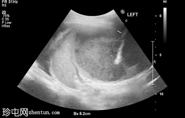
Ultrasound
从脾肿块获得的超声引导核心活检。
案例讨论
一位老年妇女因发热病入院。 腹部CT示脾脏巨大低密度肿块。 大约 2 年前的一项先前研究显示了一个小的脾肿块,该肿块尚未得到进一步研究。 目前,从肿块中迅速获得了核心活检。
脾脏(核心活检):弥漫性大 B 细胞淋巴瘤。 纤维化核心被大量非典型细胞浸润,这些细胞对 CD19、CD20、CD45 (LCA)、CD79a 和 Bcl6 呈阳性,对 CD3、CD5、CD10、CD23、细胞周期蛋白-D1 和盘角蛋白呈阴性。 Ki67指数:70-80%。 |