介绍
吐血。
患者数据
年龄:25岁
性别:女
CT
胃肠道间质瘤

Axial
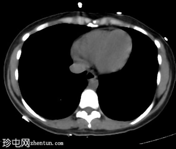
non-contrast
胃肠道间质瘤

Axial C+
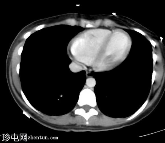
arterial phase
胃肠道间质瘤

Axial C+ portal
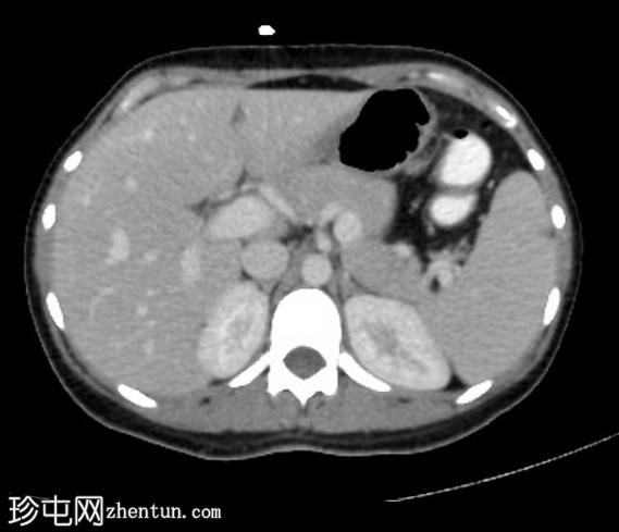
venous phase
在肝十二指肠区域可见粘膜下/粘膜延伸的大的、界限清楚的外生性、高度血管化的实性肿块病变。 它显示出内部低衰减区域的均匀增强,没有内部钙化或出血成分。
病变对邻近器官产生质量效应。
无肠梗阻。
病理
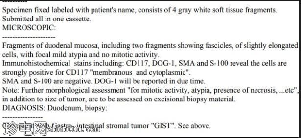
胃肠道间质瘤

案例讨论
研究结果提示十二指肠胃肠道间质瘤(GIST)。
GIST 是胃肠道最常见的间叶性肿瘤,很少发生在 40 岁之前。它们大多看起来很大,通常达到 >10 厘米,并伴有中央坏死。 然而,淋巴结肿大并不常见。
尽管肿瘤起源于粘膜下层,但很难确定大肿瘤的起源部位,因为它们可能变得非常大且外生,并可能溃疡进入粘膜。 GIST 很少引起肠梗阻,即使是大肿瘤,它也与卡尼三联征和 NF-1 相关。
参考
1. King D. The Radiology of Gastrointestinal Stromal Tumours (GIST). Cancer Imaging. 2005;5(1):150-6. doi:10.1102/1470-7330.2005.0109 - Pubmed
2. Kang H, Menias C, Gaballah A et al. Beyond the GIST: Mesenchymal Tumors of the Stomach. Radiographics. 2013;33(6):1673-90. doi:10.1148/rg.336135507 - Pubmed
3. Sandrasegaran K, Rajesh A, Rydberg J, Rushing D, Akisik F, Henley J. Gastrointestinal Stromal Tumors: Clinical, Radiologic, and Pathologic Features. AJR Am J Roentgenol. 2005;184(3):803-11. doi:10.2214/ajr.184.3.01840803 - Pubmed |