介绍
高达5–20%的胆结石病患者有胆总管(CBD)结石。仅在美国就有超过2000万人患有胆结石疾病,胆总管结石症是一个严重的全球医疗保健问题。传统上,胆石症是在“开放式”手术时代由外科医生通过开放式胆总管探查术(CBDE)专门处理的一种疾病。放射学,内窥镜检查和腹腔镜手术的进展增加了清除CBD结石的选择,同时避免了开放手术的风险[2,3]。尽管普通患者中CBDE的成功率(无论是开放式还是腹腔镜)均超过90%,但包括结石大小,胆管直径,结石数量以及是否存在解剖结构改变(周-鼻憩室,Billroth II胃空肠造口术,和Roux-en-Y配置)使某些患者的结石清除更具挑战性。本章讨论了CBDE的附件,包括胆管引流,胆道支架,球囊乳头扩张和顺行括约肌切开术。这些技术在处理更具挑战性的胆总管结石病例中可能具有重要价值。
胆漏
胆管引流允许胆管系统的外部引流,并为胆管提供通路,从而可以进行随后的胆道造影术,在某些情况下还可以进行经皮胆道干预。
T型管
T型管是长的橡胶管,末端有一条水平的短肢,位于胆管中,可引流。 T形管放置需要开腹或腹腔镜胆总管切开术。从胆管清除结石后,通过胆总管切开术将单杠置于胆管内。需要注意确保单杠的直径适合胆管的大小,并且近端(头)肢的长度合适。不合适的大横杠或长的近肢可能会导致胆道阻塞。远端肢通常长于近端肢,有些外科医生甚至更喜欢将其留得足够长,以便在Oddi括约肌进入十二指肠时将其打开。可以修整单杠的后壁,以利于管子的放置和拆卸,并改善引流效果(图9.1a-e)。胆总管切开术应使用可吸收的细缝线紧密缝合在管子周围(以防止胆汁泄漏),并注意避免在闭合过程中无意中夹住T型管。 T型管应放置至少2周,以使管周围的管道成熟。进行T管胆管造影以确认在清除T管之前的导管间隙。如果在胆道造影上看到结石,则T管束可对胆管进行经皮器械检查以清除结石。当无法通过普通导管探查作为救护措施清除CBD结石时,可以放置T型管以允许患者进行胆道引流。这可以使胆道减压,并为随后的碎石术和扩张和抽出球囊和篮的通行提供经皮通路,或者直到可以尝试进行内窥镜检查清除为止。可选地,外科医生可以使用通过胆囊管放置到总胆管(C型管)中的笔直的引流管引流胆管,并用绑带将其固定在胆囊管周围[4,5]。
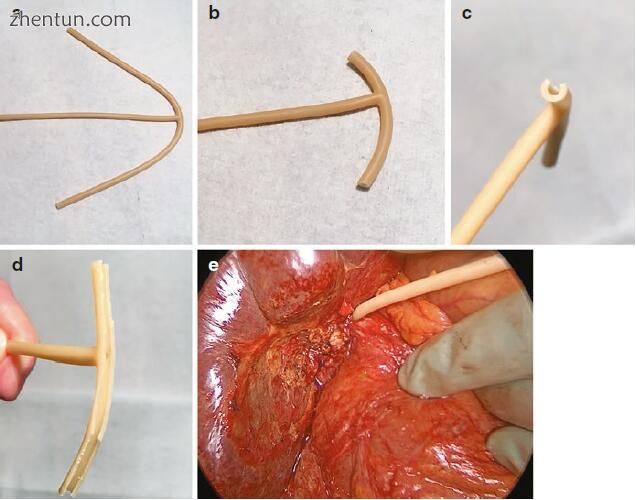
图9.1完整的T型管(a)是通过将水平肢修整成短的近端段和长的远端段(b)来形成的。可以修整(c,d)单杠的后壁,以利于管子的放置和拆卸并改善引流。 T型管可以通过胆总管切开术固定,也可以通过单独的导管切开术进行外翻,例如在Mirizzi综合征(e)的情况下,需要使用残余胆囊瓣封闭胆囊胆管瘘
尽管T管是普通导管探查的有价值的辅助手段,但初次胆总管切开术也已被证明是安全有效的[6-15]。由于对并发症的担忧,包括胆漏,胆汁性腹膜炎,体液和电解质失衡以及管移位,近年来,在T管上常规的胆总管切开术已受到严格审查。进行三项荟萃分析,比较腹腔镜导管CBDE术后的原发性导管闭合和T管上的导管闭合,与原发导管闭合相比,并发症少,手术时间短,住院时间短,成本降低到T形管的导管闭合[16-18]。 Cochrane的一项系统评价(包括6个试验和359例患者)发现,在开放性CBDE后,原发性导管闭合与T形管闭合之间的死亡率和严重发病率相似。但是,T型管组手术时间更长,住院时间更长。尽管对于简单的病例,常规的T型管使用可能不会比单纯的闭合治疗更具优势,但对于困难的病例,结石受累,术后胆道引流最重要的败血症或术后解剖结构改变的患者,它仍然是有价值的辅助手段否则将难以进入和胆道引流。
内外胆漏
胆道引流的另一种选择包括经皮放置的内外胆道引流(图9.2和9.3)。将这些尾纤导管经皮放置在肝实质中,并通过肝内胆管系统引导至肝外胆管系统进入十二指肠,在那里,尾纤部分阻止近端移入胆管。这些引流管有一个胆内段,该段具有胆汁进入胆管的侧孔,以及尾纤部分的侧孔。胆汁进入胆管内段的侧孔,然后通过尾纤段排至外袋或十二指肠。从外部夹紧管子优先使胆汁通过猪尾部分排入十二指肠。这些引流管可用于将梗阻性胆管第二段引流至胆总管结石,或用于解剖结构改变或内窥镜治疗失败的患者引流胆管损伤/泄漏。
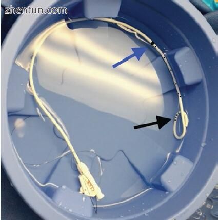
图9.2内外胆管引流。 导管具有不透射线的近端标记(蓝色箭头),标记了胆汁内引流段的最近端部分(可见侧孔)。 导管的远端部分为猪尾形状,带有十二指肠内的侧孔(黑色箭头),可防止近端迁移并允许胆汁排入十二指肠
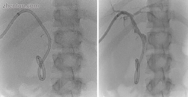
图9.3内外胆管引流。 在这种情况下放置引流管,以处理由医源性胆总管损伤引起的狭窄(在手术夹处)
还已经描述了在开放的CBDE时逆行放置内外胆管引流的外科手术技术,可以替代T型管使用[4,19]。 在开放的CBDE逆行进入肝管后,将导管通过胆管切开术推进,直到可以通过肝旁静脉触诊为止。 然后在超声引导下进一步穿刺肝实质,以避开任何主要血管。 然后通过腹壁拔出导管。 如今很少使用这种技术。
鼻胆漏
鼻胆管引流是外部胆管引流的另一种形式。在内窥镜逆行胰胆管造影术(ERCP)期间,引流管的远端通常以逆行方式置入胆管。然后,引流管的近端部分通过鼻子引出,并与重力引流管连接。鼻胆管引流管在亚洲比在美国更频繁地使用,并在特定的临床情况中起作用。例如,在严重的胆管炎需要紧急ERCP的情况下,它们允许暂时性胆道减压,以替代使用更昂贵的塑料胆道支架,从而避免在不稳定患者中遇到困难的结石时长时间尝试内镜清除结石,或者凝血病患者中括约肌切开术的更安全替代方案。鼻胆管引流通常足以在确定性治疗之前在患者稳定的情况下提供胆道引流。可以使用造影剂或什至空气进行间隔性胆管造影,以检查结石的大小和管的位置,并计划进一步的治疗,例如内镜下结石清除,普通导管探查,或在某些情况下在明确内镜下清除体外冲击波碎石术(ESWL) -ance(图9.4)。在腹腔镜或开放式跨导管CBDE期间将引流保持在适当的位置,可以关闭在胆道内胆道引流上的胆总管切开术。在这种情况下使用时,已证明与T管相比,鼻胆管引流是安全有效的。像T管一样,鼻胆管引流使外科医生可以进行术后胆道造影术,以在移开管之前确认结石清除率。鼻胆管最适合用于短期引流(以减少患者从鼻管产生的不适),与T形管不同,T管消除了在移开管之前等待尿道成熟的需要,从而缩短了患者的管持续时间[ 7、20-22]。然而,鼻胆管引流管的不利之处在于,与使用T管束不同,它们不允许经皮结石清除,并且还需要进行内窥镜检查。
胆道支架
胆管支架允许胆汁内部排入消化道。支架横穿乳头定位,其近端部分在胆管中,而远端部分在十二指肠中。胆管支架通常在内镜下放置于ERCP内,并在明确CBD结石清除,胆囊切除术后胆漏,医源性胆管损伤以及良性和恶性胆道狭窄之前进行临时引流。胆道支架也可以通过经膀胱的方法进行外科手术,以进行暂时的内部引流,通过胆总管切开术作为导管闭合的辅助手段,或作为促进术后ERCP的普通导管探查的替代方法。存在并且将讨论不同类型的胆道支架。
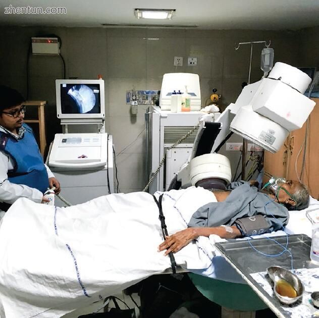
图9.4鼻胆管引流对于胆管系统的临时引流很有用。在这张照片中,鼻胆管引流到位的患者经历了外部冲击波碎石术,以分解大块的普通导管结石,从而使随后的内镜结石清除成为可能。鼻胆管引流可确保持续的胆道引流,并允许进行间隔性胆道造影以评估结石清除之前的结石进展
内窥镜支架置入
出于多种原因,可以在ERCP期间将内窥镜支架放置在CBD结石的位置。在紧急情况下,可以在胆管炎的情况下放置支架,以代替括约肌切开术(对于凝血性疾病患者)或不稳定患者的结石清除,以使胆汁持续引流并稳定患者。一旦患者康复,就可以重复进行ERCP以完成结石清除。
胆道支架在治疗难以清除的CBD结石(例如大块和多块结石)中的使用也得到了很好的描述,这是一种临时性的引流方法,可预防黄疸或胆管炎,直到所有结石可以通过后续的干预措施(包括重复的内镜检查)清除为止,经皮或手术取石。内镜下结石清除失败的患者中,有63–96%的患者在支架置入成功后的3– 6个月重复ERCP清除结石。胆道支架本身的存在被认为可导致结石尺寸减小,并可能导致结石碎裂或软化,这可以促进碎石术和用气球取出结石[24-27]。相反,添加药理学的剂(如熊去氧胆酸)似乎并没有减少结石的大小,增加结石的碎裂或促进随后的结石提取[28,29]。支架对内镜医师也有帮助,因为它们的存在会促进乳头的重复插管,以及在深层胆管插管困难的情况下(例如,存在胆道狭窄时)。
与临时使用相反,对于那些具有高手术风险的CBD结石患者(例如老年人或多种合并症患者,内镜下导管清除困难或失败),建议将支架作为括约肌切开术的长期辅助手段[30–36]。支架可使胆管减压,从而消除黄疸并预防胆管炎,并且可以在体弱的患者或寿命有限的患者中用作确定性措施。一项对201例不适合内镜摘除的结石患者进行了括约肌切开术和7 F双尾钉支架置入术的研究发现,中位支架通畅性接近5年,复发性胆管炎的发生率为7.4%,复发性黄疸的发生率为18.5%。在选择长期支架治疗的策略时,一些内镜医师倾向于常规更换支架以防止阻塞和胆管炎,而另一些内镜医师则认为可以进行安全的长期治疗,而无需常规支架更换。使用两个或多个塑料支架作为胆总管结石的最终治疗方法可能有助于减轻支架闭塞的风险[28,36–39]。
塑料胆道支架
临时性塑料支架是治疗胆总管结石最常用的支架。这些支架使用不同的固定系统来防止支架迁移,包括瓣和尾纤末端。装有瓣的支架是直的塑料管,其利用瓣将支架近端和远端固定在适当的位置,以分别防止意外的逆行或顺行迁移。支架的尺寸从7 F到12 F不等,长度从5到15 cm不等。将支架放置在胆管中,使含瓣的远端伸入十二指肠(图9.5)。尾纤支架利用环形挠性端将支架固定在适当位置(图9.6)。胆汁通过毛细作用流过支架以及支架周围。塑料支架最多可以保留3个月,以免发生阻塞和随后发生胆管炎的风险。
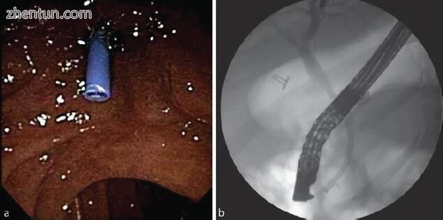
图9.5(a)瓣固定塑料支架的内窥镜视图。 (b)皮瓣固定塑料支架的透视图
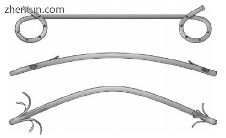
图9.6塑料胆道支架(Bruno MJ。Ch 51.恶性胰胆管梗阻的缓解。
金属胆道支架
金属支架由金属合金组成,一旦部署在胆道系统中,金属合金会膨胀到预定形状。金属支架类型包括未覆盖的自膨胀金属支架(SEMS),部分覆盖的SEMS和完全覆盖的SEMS。裸露的SEMS不能去除,因此,由于组织向内生长会阻止安全去除,因此它们的使用应仅限于减轻恶性胆道狭窄。覆膜支架由覆盖有各种材料的金属格子组成,以防止组织或肿瘤向内生长。这些覆盖的支架是可移动的,因此可用于良性狭窄或困难的普通导管结石。已经描述了使用这些支架治疗CBD结石,其结果类似于塑料支架,其并发症包括支架移入或移出胆管,胰腺炎,穿孔和出血[41,42]。目前尚不清楚,与较便宜的塑料支架相比,在结石清除率或并发症方面,较困难的胆总管结石病例使用较昂贵的有盖SEMS是否能带来任何益处。表9.1比较了不同类型的胆道支架。
手术支架放置
胆道支架也可以在开放式或腹腔镜CBDE手术中放置。很好地描述了在塑料胆道支架(猪尾型和直瓣固定型支架)上进行胆总管切开术的闭合过程。可以通过胆管切开术放置支架,或通过导丝通过胆囊管顺行放置支架,然后闭合胆总管切开术[7,43-50]。与通过T管闭合胆总管切开术相比,支架置入与手术时间短和住院时间短有关[51-54]。为此目的使用支架的缺点是需要用于支架取出的术后上内窥镜检查。一些外科医生通过移除近端锚固件(皮瓣或猪尾环)并用可快速吸收的缝合线材料代替它来修改支架,以允许支架自发地和有意地通过消化道向远侧迁移,从而无需术后进行内镜检查[54–57]。对于解剖结构改变的患者(Billroth II或Roux-en-Y胃空肠吻合术),ERCP可能极具挑战性,胆总管切开或胆道引流(T管或C管)的初次闭合可能比a更为合适。支架在这种情况下。对于小口径胆管患者,通过胆道支架进行导管闭合可能比不采用支架的T管放置或导管闭合更好,以避免狭窄形成。但是,缺乏支持这种做法的强大数据。表9.2比较了在CBDE期间关闭胆总管切开术的不同方法。
支架相关并发症
使用胆道支架是安全的,但可能会引起并发症。最常见的包括支架阻塞,导致黄疸,肝功能酶升高和胆管炎。塑料支架的支架迁移(近端或远端)可能发生在3%到10%之间。近端移入胆管系统使内窥镜切除具有挑战性。远端迁移到胃肠道通常会导致支架完全通过而不会出现任何问题[58,59]。胆道支架迁移[60-62]或通过胆道支架腐蚀支架[63,64]很少导致肠穿孔。
球囊乳头扩张
内镜下球囊乳头扩张术(EBPD)于1982年首次描述,已被用作内镜括约肌切开术的替代方案和辅助措施,以方便地清除CBD结石[65-70]。与括约肌切开术相比,EBPD(至少在理论上而言)保持了Oddi括约肌的完整性,并可以防止十二指肠内容物回流到胆管系统中而引起的长期并发症。内窥镜球囊扩张术在具有挑战性的解剖结构患者(例如Billroth II胃空肠造口术)以及凝血病患者中尤其有用[23,71–73]。
EBPD通过在深部插管后将一个较大的径向扩张的球囊通入胆管来进行。 将球囊充气(最大直径为20 mm)一段时间(30 s至5分钟),导致Oddi括约肌伸展。 基于所施加的大气压,膨胀球囊可靠地膨胀至预定直径。 使用手动压力泵可以将球囊的充气和放气控制到所需的直径(图9.7)。 如果最初的内镜括约肌切开术不足以取回大块结石,则从EBPD进行的额外拉伸可以促进结石的清除。 尽管最初犹豫不决,但多项研究表明,内镜括约肌切开术后的EBPD是安全的,包括某些高危患者(例如壶腹周围憩室的患者)[75-77]。

图9.7内窥镜球囊乳头扩张术。 (a)充气气球的内窥镜视图。 (b)充气气球的透视图。 (c)成功扩张后扩张乳头的内窥镜视图
该技术是从内窥镜领域借来的,并用作经皮和外科CBDE(腹腔镜或开放式)的辅助手段[78-80]。扩张球囊在荧光镜的引导下通过跨囊或跨导方法顺行穿过Oddi括约肌。扩张完成后,使用高压冲洗(冲洗)更可能导致结石移位进入十二指肠。或者,可以使用提取球囊或胆道镜将结石通过扩张的括约肌向远端推入十二指肠。当球囊位于结石远端的括约肌处时,应小心谨慎,同时撤回放气的球囊,以防止结石近端移入肝管。
EBPD清除结石的成功率超过90%,与内镜括约肌切开术清除大结石的成功率相当。该技术的10%并发症发生率也类似于内窥镜括约肌切开术。然而,内镜括约肌切开术后出血更为普遍,而球囊扩张术后胰腺炎似乎更为常见。接受EBPD的患者发生ERCP后胰腺炎发生率增加的背后机制尚不清楚。一项回顾性研究表明,在内镜下进行球囊-乳头扩张术时,高淀粉血症的发生率为30%,而经皮进行的则为7%。球囊充气的持续时间可能对胰腺炎的发生率有影响。研究表明,在没有进行括约肌切开术的情况下进行EBPD时,气囊充气的持续时间较短(<5分钟,≥5分钟)会导致较高的胰腺炎发生率(18%对0%)。假设较短的球囊扩张持续时间可能导致Oddi纤维括约肌的完全破坏不完全,从而导致胰管肿胀,痉挛和术后阻塞,从而导致胰腺炎。内镜下EBPD而非经皮EBPD所见的胰腺炎发生率更高,这表明其他因素(例如,包括碎石术导管和篮在内的较大器械的通过而不是球囊扩张本身)可能导致胰腺炎的发生率更高。 EBPD的另一种并发症包括胆管损伤。因此,所用球囊的尺寸不应超过胆管的直径。
括约肌切开术
一体化括约肌切开术是一种术中选择,可用于在术中ERCP期间促进深层胆管插管或作为普通导管探查的辅助手段。外科医生顺行地将导丝穿过囊状导管穿过乳头并进入十二指肠。然后将括约肌切开器通过导线并用于产生括约肌切开术。这应在侧视十二指肠镜的内窥镜引导下进行。在与胰管位置相对的乳头的11点或12点位置进行适当大小的括约肌切开术。然后从上方冲洗或将总管结石冲洗或推入十二指肠(图9.8a–c)。经皮和腹腔镜均已描述了该技术[5,84–86]。此技术类似于会合过程,在该过程中,外科医生使金属丝顺行穿过囊性导管并穿过乳头,这样ERCP操作员就可以将金属丝圈套起来并获得用于逆行括约肌切开术的金属丝通道。然后,ERCP操作员以常规方式进行石材提取。与会合手术相比,已发现顺行括约肌切开术与更短的手术时间和相当的结石取出率和并发症发生率相关。
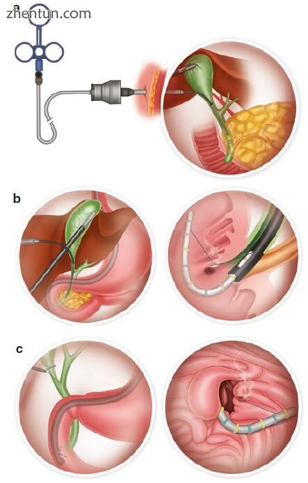
图9.8腹腔镜顺行括约肌切开术。 (a)使用导引鞘将标准的内窥镜括约肌切开术通过右上象限5.0 mm套管插入,以最大程度地减少气体泄漏。 (b)左:十二指肠镜位于壶腹的正对面,因此可以在直视下将括约肌切开器引导到正确的位置。右:括约肌弓形器被弯成弓形,露出切割丝,并进行操纵直到其位于12点钟的位置。 (c)左:可使用导丝促进括约肌刀穿过壶腹。右:凝结和切割电流的混合物用于将括约肌划分到十二指肠的第一个横向折叠处
结论
胆总管结石症疑难病例的处理需要对不同方法及其优缺点的充分了解。具有高手术风险和解剖结构改变的患者以及结石受累的患者可从胆总管探查的辅助手段中受益,包括胆道引流,胆道支架,球囊乳头扩张和顺行括约肌切开术。适当使用这些不同的方法(无论是内窥镜检查,经皮手术,外科手术或方法的组合)都可以在最小化并发症的同时,优化胆总管结石的处理。
参考文献
Choledocholithiasis Comprehensive Surgical Management
1.Everhart JE, Khare M, Hill M, Maurer KR. Prevalence and ethnic differences in gallbladder disease in the United States. Gastroenterology. 1999;117(3):632–9.
2.Hazey JW. Historical perspective and treatments for common bile duct stones. In: Hazey JW, Conwell DL, Guy GE, editors. Multidisciplinary management of common bile duct stones. Cham: Springer International Publishing; 2016. p. 1–4.
3.Khadra H, Jackson T, Marks J. Management of surgical complications. In: Hazey JW, Conwell DL, Guy GE, editors. Multidisciplinary management of common bile duct stones. Cham: Springer International Publishing; 2016. p. 129–44.
4.Hotta T, Taniguchi K, Kobayashi Y, Johata K, Sahara M, Naka T, et al. Biliary drainage tube evaluation after common bile duct exploration for choledocholithiasis. Hepatogastroenterology. 2003;50(50):315–21.
5.Ponsky JL, Heniford BT, Gersin K. Choledocholithiasis: evolving intraoperative strategies. Am Surg. 2000;66(3):262–8.
6.Ambreen M, Shaikh AR, Jamal A, Qureshi JN, Dalwani AG, Memon MM. Primary closure versus T-tube drainage after open choledochotomy. Asian J Surg. 2009;32(1):21–5.
7.Chen C-C, Wu S-D, Tian Y, Zeng X-T, Siwo EA, Xian G-Z. The fading role of T-tube in lapa-roscopic choledochotomy: primary choledochorrhaphy and over pigtail j and endonasobiliary drainage tubes. J Laparoendosc Adv Surg Tech A. 2010;20(10):807–11.
8.Decker G, Borie F, Millat B, Berthou JC, Deleuze A, Drouard F, et al. One hundred lap-aroscopic choledochotomies with primary closure of the common bile duct. Surg Endosc. 2003;17(1):12–8.
9.El-Geidie AAR. Is the use of T-tube necessary after laparoscopic choledochotomy? J Gastrointest Surg. 2010;14(5):844–8.
10.Gurusamy KS, Samraj K. Primary closure versus T-tube drainage after open common bile duct exploration. Cochrane Database Syst Rev. 2007;(1):CD005640.
11.Saeed N, Nasir MT, Khan MI, Channa GA. Choledochorraphy (primary repair) versus t-tube drainage after open choledochotomy. J Ayub Med Coll Abbottabad JAMC. 2012;24(1):18–20.
12.Sorensen VJ, Buck JR, Chung SK, Fath JJ, Horst HM, Obeid FN. Primary common bile duct closure following exploration: an effective alternative to routine biliary drainage. Am Surg. 1994;60(6):451–4.
13.Zhu QD, Tao CL, Zhou MT, Yu ZP, Shi HQ, Zhang QY. Primary closure versus T-tube drain-age after common bile duct exploration for choledocholithiasis. Langenbecks Arch Surg. 2011;396(1):53–62.
14.Zhang H-W, Chen Y-J, Wu C-H, Li W-D. Laparoscopic common bile duct exploration with pri-mary closure for management of choledocholithiasis: a retrospective analysis and comparison with conventional T-tube drainage. Am Surg. 2014;80(2):178–81.
15.Ahmed I, Pradhan C, Beckingham IJ, Brooks AJ, Rowlands BJ, Lobo DN. Is a T-tube neces-sary after common bile duct exploration? World J Surg. 2008;32(7):1485–8.
16.Podda M, Polignano FM, Luhmann A, Wilson MSJ, Kulli C, Tait IS. Systematic review with meta-analysis of studies comparing primary duct closure and T-tube drainage after laparoscopic common bile duct exploration for choledocholithiasis. Surg Endosc. 2016;30(3):845–61.
17.Wu X, Yang Y, Dong P, Gu J, Lu J, Li M, et al. Primary closure versus T-tube drainage in laparoscopic common bile duct exploration: a meta-analysis of randomized clinical trials. Langenbecks Arch Surg. 2012;397(6):909–16.
18.Yin Z, Xu K, Sun J, Zhang J, Xiao Z, Wang J, et al. Is the end of the T-tube drainage era in laparoscopic choledochotomy for common bile duct stones is coming? A systematic review and meta-analysis. Ann Surg. 2013;257(1):54–66.
19.Kusano T, Isa T, Shimoji H, Miyazato H, Muto Y, Furukawa M. Results of retrograde tran-shepatic biliary drainage after a common bile duct exploration for choledocholithiasis. Hepatogastroenterology. 1999;46(29):2776–80.
20.Otani K, Ueki T, Matsumura K, Maruo T, Minoda R, Otsuka Y, et al. Comparison between endoscopic biliary stenting and nasobiliary drainage in patients with acute cholangitis due to choledocholithiasis: is endoscopic biliary stenting useful? Hepatogastroenterology. 2015;62(139):558–63.
21.Wani MA, Chowdri NA, Naqash SH, Parray FQ, Wani RA, Wani NA. Closure of the com-mon duct-endonasobiliary drainage tubes vs. t tube: a comparative study. Indian J Surg. 2010;72(5):367–72.
22.Zhang H-F, Hu S-Y, Zhang G-Y, Wang K-X, Chen B, Li B. Laparoscopic primary choledo-chorrhaphy over endonasobiliary drainage tubes. Surg Endosc. 2007;21(11):2115–7.
23.Pearl J. Techniques of endoscopic retrograde cholangiopancreatography. In: Marks JM, Dunkin BJ, editors. Principles of flexible endoscopy for surgeons. New York: Springer; 2013. p. 215–26.
24.Aslan F, Arabul M, Celik M, Alper E, Unsal B. The effect of biliary stenting on difficult com-mon bile duct stones. Przeglad Gastroenterol. 2014;9(2):109–15.
25.Horiuchi A, Nakayama Y, Kajiyama M, Kato N, Kamijima T, Graham DY, et al. Biliary stent-ing in the management of large or multiple common bile duct stones. Gastrointest Endosc. 2010;71(7):1200–3.e2.
26.Fan Z, Hawes R, Lawrence C, Zhang X, Zhang X, Lv W. Analysis of plastic stents in the treat-ment of large common bile duct stones in 45 patients. Dig Endosc. 2011;23(1):86–90.
27.Sharma SS, Jhajharia A, Maharshi S. Short-term biliary stenting before mechanical lithotripsy for difficult bile duct stones. Indian J Gastroenterol. 2014;33(3):237–40.
28.Lee TH, Han J-H, Kim HJ, Park SM, Park S-H, Kim S-J. Is the addition of choleretic agents in multiple double-pigtail biliary stents effective for difficult common bile duct stones in elderly patients? A prospective, multicenter study. Gastrointest Endosc. 2011;74(1):96–102.
29.Katsinelos P, Kountouras J, Paroutoglou G, Chatzimavroudis G, Zavos C. Combination of endoprostheses and oral ursodeoxycholic acid or placebo in the treatment of difficult to extract common bile duct stones. Dig Liver Dis. 2008;40(6):453–9.
30.Ang TL, Fock KM, Teo EK, Chua TS, Tan J. An audit of the outcome of long-term biliary stenting in the treatment of common bile duct stones in a general hospital. J Gastroenterol. 2006;41(8):765–71.
31.Hong W, Zhu Q, Huang Q. Endoscopic sphincterotomy plus endoprostheses in the treatment of large or multiple common bile duct stones. Dig Endosc. 2011;23(3):240–3.
32.Horiuchi A, Kajiyama M, Ichise Y, Tada H, Fujiwara N, Tanaka N. Biliary stenting as alterna-tive therapy to stone clearance in elderly patients with bile duct stones. Acta Gastro-Enterol Belg. 2014;77(3):297–301.
33.Li KW, Zhang XW, Ding J, Chen T, Wang J, Shi WJ. A prospective study of the efficacy of endoscopic biliary stenting on common bile duct stones. J Dig Dis. 2009;10(4):328–31.
34.Peters R, Macmathuna P, Lombard M, Karani J, Westaby D. Management of common bile duct stones with a biliary endoprosthesis. Report on 40 cases. Gut. 1992;33(10):1412–5.
35.Yang J, Peng J, Chen W. Endoscopic biliary stenting for irretrievable common bile duct stones: indications, advantages, disadvantages, and follow-up results. Surg J R Coll Surg Edinb Irel. 2012;10(4):211–7.
36.Slattery E, Kale V, Anwar W, Courtney G, Aftab AR. Role of long-term biliary stenting in choledocholithiasis. Dig Endosc. 2013;25(4):440–3.
37.Di Giorgio P, Manes G, Grimaldi E, Schettino M, D’Alessandro A, Di Giorgio A, et al. Endoscopic plastic stenting for bile duct stones: stent changing on demand or every 3 months. A prospective comparison study. Endoscopy. 2013;45(12):1014–7.
38.Maxton DG, Tweedle DE, Martin DF. Stenting for choledocholithiasis: temporizing or thera-peutic? Am J Gastroenterol. 1996;91(3):615–6.
39.Ye X, Huai J, Sun X. Effectiveness and safety of biliary stenting in the management of difficult common bile duct stones in elderly patients. Turk J Gastroenterol. 2016;27(1):30–6.
40.Bruno MJ. Palliation of malignant pancreaticobiliary obstruction. In: Ginsberg GG, Gostout CJ, Kochman ML, Norton ID, editors. Clinical gastrointestinal endoscopy. St. Louis: Elsevier Saunders; 2011. p. 706–20.
41.Bakhru MR, Kahaleh M. Expandable metal stents for benign biliary disease. Gastrointest Endosc Clin N Am. 2011;21(3):447–62, viii.
42.Cerefice M, Sauer B, Javaid M, Smith LA, Gosain S, Argo CK, et al. Complex biliary stones: treatment with removable self-expandable metal stents: a new approach (with videos). Gastrointest Endosc. 2011;74(3):520–6.
43.Dietrich A, Alvarez F, Resio N, Mazza O, de Santibañes E, Pekolj J, et al. Laparoscopic man-agement of common bile duct stones: transpapillary stenting or external biliary drainage? JSLS. 2014;18(4):pii: e2014.00277.
44.Fujii M, Okino M, Fujioka K, Yamashita K, Hamano K. Placement of a double-pigtail ERBD tube after primary closure of the common bile duct in three patients with choledocholithiasis. J Hepatobiliary Pancreat Surg. 2005;12(5):386–90.
45.Isla AM, Griniatsos J, Karvounis E, Arbuckle JD. Advantages of laparoscopic stented choledo-chorrhaphy over T-tube placement. Br J Surg. 2004;91(7):862–6.
46.Kuroki T, Tajima Y, Tsuneoka N, Kitasato A, Adachi T, Kosaka T, et al. Placement of a plastic biliary stent tube with primary closure of the common bile duct after laparoscopic common bile duct exploration. Hepatogastroenterology. 2010;57(102–103):1034–6.
47.Lee JS, Yoon YC. Laparoscopic common bile duct exploration using V-Loc suture with inser-tion of endobiliary stent. Surg Endosc. 2016;30(6):2530–4.
48.Wei Q, Hu H-J, Cai X-Y, Li L-B, Wang G-Y. Biliary drainage after laparoscopic choledo-chotomy. World J Gastroenterol. 2004;10(21):3175–8.
49.Bandyopadhyay SK, Khanna S, Sen B, Tantia O. Antegrade common bile duct (CBD) stenting after laparoscopic CBD exploration. J Minimal Access Surg. 2007;3(1):19–25.
50.Pérez G, Escalona A, Jarufe N, Ibáñez L, Viviani P, García C, et al. Prospective randomized study of T-tube versus biliary stent for common bile duct decompression after open choledo-cotomy. World J Surg. 2005;29(7):869–72.
51.Lyon M, Menon S, Jain A, Kumar H. Use of biliary stent in laparoscopic common bile duct exploration. Surg Endosc. 2015;29(5):1094–8.
52.Tang CN, Tai CK, Ha JPY, Tsui KK, Wong DCT, Li MKW. Antegrade biliary stenting versus T-tube drainage after laparoscopic choledochotomy – a comparative cohort study. Hepatogastroenterology. 2006;53(69):330–4.
53.Xin Y, Zhu X, Wei Q, Cai X, Wang X, Huang D. Comparison of quality of life between two biliary drainage procedures in laparoscopic common bile duct exploration. Hepatogastroenterology. 2007;54(74):331–3.
54.Xu Y, Dong C, Ma K, Long F, Jiang K, Shao P, et al. Spontaneously removed biliary stent drainage versus T-tube drainage after laparoscopic common bile duct exploration. Medicine (Baltimore). 2016;95(39):e5011.
55.Huang J, Zhu J. Spontaneously removed endobiliary J stent drainage after laparoscopic com-mon bile duct exploration. Surg Endosc. 2009;23(6):1398–402.
56.Kim EK, Lee SK. Laparoscopic treatment of choledocholithiasis using modified biliary stents. Surg Endosc. 2004;18(2):303–6.
57.Tian MG, Shi WJ, Zhong CJ, Zhang XW, Chen TM. Laparoscopic treatment of cho-ledocholithiasis with novel self-releasing biliary stent. J Laparoendosc Adv Surg Tech A. 2009;19(3):405–8.
58.Cerisoli C, Diez J, Giménez M, Oria M, Pardo R, Pujato M. Implantation of migrated biliary stents in the digestive tract. HPB. 2003;5(3):180–2.
59.Katsinelos P, Kountouras J, Paroutoglou G, Chatzimavroudis G, Paikos D, Zavos C, et al. Migration of plastic biliary stents and endoscopic retrieval: an experience of three referral centers. Surg Laparosc Endosc Percutan Tech. 2009;19(3):217–21.
60.Chittleborough TJ, Mgaieth S, Kirkby B, Zakon J. Remove the migrated stent: sigmoid colon perforation from migrated biliary stent. ANZ J Surg. 2016;86(11):947–8.
61.El Zein MH, Kumbhari V, Tieu A, Saxena P, Messallam AA, Azola A, et al. Duodenal perfora-tion as a consequence of biliary stent migration can occur regardless of stent type or duration. Endoscopy. 2014;46(Suppl 1):E281–2.
62.Zeb F, Kevans D, Muir K, Courtney G, Tadros E, Aftab A. Duodenal impaction/perforation of a biliary stent – a rare complication in the management of choledocholithiasis. J Gastrointest Liver Dis JGLD. 2009;18(3):391–2.
63.Dundee PE, Franzi SJ, Thomas PR. Unique complication of a biliary stent: erosion through the common hepatic duct wall. ANZ J Surg. 2007;77(3):194.
64.Sinha S, Wardle A, Kalidindi V, Banga N, Stoker DL. Erosion and perforation of the biliary tree by plastic biliary endoprostheses. Endoscopy. 2010;42(9):760–3.
65.Chung JW, Chung JB. Endoscopic papillary balloon dilation for removal of choledocholi-thiasis: indications, advantages, complications, and long-term follow-up results. Gut Liver. 2011;5(1):1–14.
66.Nagashima I, Takada T, Shiratori M, Inaba T, Okinaga K. Percutaneous transhepatic papillary balloon dilation as a therapeutic option for choledocholithiasis. J Hepatobiliary Pancreat Surg. 2004;11(4):252–4.
67.Tsai T-J, Lai K-H, Lin C-K, Chan H-H, Wang E-M, Tsai W-L, et al. Role of endoscopic pap-illary balloon dilation in patients with recurrent bile duct stones after endoscopic sphincter-otomy. J Chin Med Assoc JCMA. 2015;78(1):56–61.
68.Watanabe H, Hiraishi H, Koitabashi A, Sasai T, Kanke K, Oinuma T, et al. Endoscopic pap-illary balloon dilation for treatment of common bile duct stones. Hepatogastroenterology. 2004;51(57):652–7.
69.Zippi M, De Felici I, Pica R, Traversa G, Occhigrossi G. Endoscopic papillary balloon dilation for difficult common bile duct stones: our experience. World J Clin Cases. 2013;1(1):19–24.
70.Testoni PA, Mariani A, Aabakken L, Arvanitakis M, Bories E, Costamagna G, et al. Papillary cannulation and sphincterotomy techniques at ERCP: European Society of Gastrointestinal Endoscopy (ESGE) Clinical Guideline. Endoscopy. 2016;48(7):657–83.
71.Shakhatreh MH, Groce JR. Gastroenterologic treatment and outcomes. In: Hazey JW, Conwell DL, Guy GE, editors. Multidisciplinary management of common bile duct stones. Cham: Springer International Publishing; 2016. p. 85–91.
72.Baron TH, Harewood GC. Endoscopic balloon dilation of the biliary sphincter compared to endoscopic biliary sphincterotomy for removal of common bile duct stones during ERCP: a metaanalysis of randomized, controlled trials. Am J Gastroenterol. 2004;99(8):1455–60.
73.Jang HW, Lee KJ, Jung MJ, Jung JW, Park JY, Park SW, et al. Endoscopic papillary large bal-loon dilatation alone is safe and effective for the treatment of difficult choledocholithiasis in cases of Billroth II gastrectomy: a single center experience. Dig Dis Sci. 2013;58(6):1737–43.
74.Lai K-H, Chan H-H, Tsai T-J, Cheng J-S, Hsu P-I. Reappraisal of endoscopic papillary bal-loon dilation for the management of common bile duct stones. World J Gastrointest Endosc. 2015;7(2):77–86.
75.Kim KH, Kim TN. Endoscopic papillary large balloon dilation in patients with periampullary diverticula. World J Gastroenterol. 2013;19(41):7168–76.
76.Poincloux L, Rouquette O, Privat J, Gorce D, Abergel A, Dapoigny M, et al. Large-balloon dilation of the sphincter of Oddi after sphincterotomy or infundibulotomy to extract large calculi or multiple common bile duct stones without using mechanical lithotripsy. Scand J Gastroenterol. 2013;48(2):246–51.
77.Sakai Y, Tsuyuguchi T, Sugiyama H, Nishikawa T, Kurosawa J, Saito M, et al. Endoscopic sphincterotomy combined with large balloon dilation for removal of large bile duct stones. Hepatogastroenterology. 2013;60(121):58–64.
78.Shirai N, Hanai H, Kajimura M, Kataoka H, Yoshida K, Nakagawara M, et al. Successful treat-ment of percutaneous transhepatic papillary dilation in patients with obstructive jaundice due to common bile duct stones after Billroth II gastrectomy: report of two emergent cases. J Clin Gastroenterol. 2000;30(1):91–3.
79.Ozcan N, Kahriman G, Mavili E. Percutaneous transhepatic removal of bile duct stones: results of 261 patients. Cardiovasc Intervent Radiol. 2012;35(4):890–7.
80.Kumar A, Martinez JM. Surgical treatment and outcomes. In: Hazey JW, Conwell DL, Guy GE, editors. Multidisciplinary management of common bile duct stones. Cham: Springer International Publishing; 2016. p. 93–100.
81.Masoni L, Mari FS, Pietropaolo V, Onorato M, Meucci M, Brescia A. Laparoscopic treatment for unsuspected common bile duct stones by transcystic sphincter of Oddi pneumatic balloon dilation and pressure-washing technique. World J Surg. 2013;37(6):1258–62.
82.Shen YH, Yang LQ, Yao YL, Wang L, Zhang YY, Cao J, He QB, Zou XP, Li YH. Dilation time in endoscopic papillary balloon dilation for common bile duct stones. Surg Laparosc Endosc Percutan Tech. 2017;27(5):351–5.
83.Seo YR, Moon JH, Choi HJ, Kim DC, Lee TH, Cha S-W, et al. Papillary balloon dilation is not itself a cause of post-endoscopic retrograde cholangiopancreatography pancreatitis; results of anterograde and retrograde papillary balloon dilation. J Gastroenterol Hepatol. 2013;28(8):1416–21.
84.Born P, Sandschin W, Rsch T. Percutaneous antegrade sphincterotomy under endoscopic ret-rograde control: report of two cases. Endoscopy. 2002;34(6):512–3.
85.DePaula AL, Hashiba K, Bafutto M, Zago R, Machado MM. Laparoscopic antegrade sphinc-terotomy. Surg Laparosc Endosc. 1993;3(3):157–60.
86.Hunter JG, Pham TH. Laparoscopic cholecystectomy, intraoperative cholangiography, and common bile duct exploration. In: Fischer JE, editor. Mastery of surgery. Philadelphia: Lippincott Williams and Wilkins; 2006. p. 1265–77.
87.Tekin A, Ogetman Z, Altunel E. Laparoendoscopic “rendezvous” versus laparoscopic ante-grade sphincterotomy for choledocholithiasis. Surgery. 2008;144(3):442–7. |