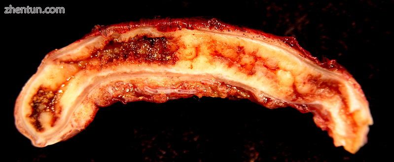
阑尾炎是阑尾炎症。[2] 症状通常包括右下腹痛,恶心,呕吐和食欲下降[2]。 然而,大约40%的人没有这些典型症状。[2] 阑尾破裂的严重并发症包括腹壁内层和脓毒症的广泛且疼痛的炎症[3]。
阑尾炎是由于阑尾中空部分堵塞造成的[10]。 这最常见的原因是由粪便制成的钙质“石头”。[6] 来自病毒感染的炎症淋巴组织,寄生虫,胆结石或肿瘤也可能导致堵塞。[6] 这种堵塞导致阑尾压力增加,阑尾组织血流量减少,阑尾内部细菌生长导致炎症[6] [11]。 炎症,阑尾血流量减少和阑尾膨胀导致组织损伤和组织死亡[12]。 如果这个过程没有得到治疗,阑尾可能爆裂,释放细菌进入腹腔,导致并发症增加。[12] [13]
阑尾炎的诊断主要基于患者的体征和症状[11]。 如果诊断不明确,密切观察,医学影像和实验室检查可能会有所帮助[4]。 使用的两种最常见的成像测试是超声和计算机断层扫描(CT扫描)。[4] CT检查显示比超声检查更准确的检测急性阑尾炎[14]。 然而,由于CT扫描的辐射暴露风险,超声可能首选作为儿童和孕妇的首次成像检查[4]。
急性阑尾炎的标准治疗是手术切除阑尾。[6] [11] 这可以通过腹部的开放切口(剖腹手术)或通过相机(腹腔镜)的一些较小的切口来完成。 手术降低了与阑尾破裂相关的副作用或死亡风险[3]。 在某些非破裂性阑尾炎病例中,抗生素可能同样有效[7]。 这是导致严重腹痛的最常见和最重要的原因之一。 2015年,约有1160万例阑尾炎发生,导致约50,100人死亡。[8] [9] 在美国,阑尾炎是需要手术的突发腹痛的最常见原因[2]。 美国每年有超过300,000名阑尾炎患者手术切除阑尾。[15] 雷金纳德菲茨被认为是在1886年第一个描述该病情的人。[16]
目录
1体征和症状
2原因
3诊断
3.1临床
3.2血液和尿液检测
3.3成像
3.4评分系统
3.5病理学
3.6鉴别诊断
4管理
4.1疼痛
4.2手术
5预后
6流行病学
7参考文献
体征和症状
Location of the appendix in the digestive system
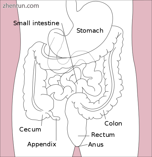
附件在消化系统中的位置
急性阑尾炎的表现包括腹痛,恶心,呕吐和发烧。当阑尾变得更加肿胀并且发炎时,它开始刺激相邻的腹壁。这导致疼痛局部化到右下象限。三岁以下的儿童可能不会看到这种典型的疼痛迁移。这种疼痛可以通过体征引起,并且可以是严重的。体征包括右侧髂窝局部发现。腹壁对温和的压力(触诊)非常敏感。下腹部突然发生深部压痛(反跳痛)时疼痛剧烈。如果阑尾位于盲肠后方(局部位于盲肠后面),则右下腹部的深部压力可能无法引起压痛(无声附件)。这是因为盲肠与气体扩张,保护发炎的阑尾免受压力。同样,如果阑尾完全位于骨盆内,通常完全没有腹部僵硬。在这种情况下,数字直肠检查可引起直肠包囊的压痛。咳嗽在这个区域引起点压痛(McBurney的观点),历史上称为Dunphy's征。
原因
急性阑尾炎似乎是阑尾主要阻塞的最终结果。[17] [10]一旦发生这种阻塞,阑尾就会充满粘液和肿胀。粘液持续产生导致阑尾内腔和壁内的压力增加。增加的压力导致小血管的血栓形成和闭塞,以及淋巴流动的停滞。此时很少发生自发恢复。随着血管闭塞的进展,阑尾变成缺血性坏死。随着细菌开始通过垂死的墙壁泄漏,脓液在阑尾内和周围形成(化脓)。最终的结果是引起腹膜炎的阑尾破裂(“爆发性阑尾”),这可能导致败血症并最终导致死亡。这些事件是缓慢演变的腹痛和其他常见症状的原因。[12]
致病因子包括牛黄,异物,创伤,肠蠕虫,淋巴结炎,最常见的是钙化的粪便沉积物,称为阑尾或fecoliths [18] [19]。阻碍粪便的发生已经引起了人们的关注,因为它们在阑尾炎患者中的发病率高于发展中国家[20]。此外,阑尾粪便通常与复杂的阑尾炎相关[21]。正如对急性阑尾炎患者每周排便次数少于健康对照者所证实的,粪便淤滞和停滞可能起到一定的作用[19] [22]。
在阑尾中发现粪便被认为是由于结肠中的右侧粪便滞留储库和延长的运输时间。 然而,在随后的研究中没有观察到延长的运送时间[23]。 从流行病学资料来看,有报道说憩室病和腺瘤性息肉不详,结肠癌在社区中极为罕见,可免除阑尾炎[24] [25]。 已证明急性阑尾炎先于结肠和直肠中的癌症[26]。 几项研究提供了低纤维摄入量参与阑尾炎发病机制的证据。[27] [28] [29] 膳食纤维的这种低摄入量与右侧粪便储存库的出现以及膳食纤维减少通行时间的事实一致[30]。
诊断
Appendicitis as seen on CT imaging
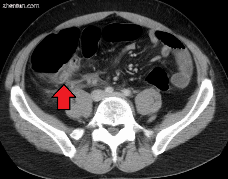
在CT成像上看到的阑尾炎
诊断基于病史(症状)和体检,如果需要,可以通过升高中性白细胞和成像研究来支持。 (嗜中性粒细胞是对细菌感染有反应的主要白细胞。)历史分为两类,典型的和非典型的。
典型的阑尾炎包括在肚脐区域开始伴有相关的厌食,恶心或呕吐的数小时的全身腹痛。然后疼痛“局部化”到柔软度增加的右下腹部。有可能疼痛可能局部位于左侧下象限位点转位的患者。疼痛,厌食,白细胞增多和发热的组合是经典的。
非典型病史缺乏这种典型的进展,可能包括右下腹部疼痛作为初始症状。腹膜刺激(腹壁内侧)可导致运动疼痛增加或颠簸,例如超速。[31]非典型病史通常需要使用超声或CT扫描进行成像[32]。
临床
Aure-Rozanova征:右手指触诊增加疼痛Petit三角(可以是积极的Shchetkin-Bloomberg's)[33]
Bartomier-Michelson的标志:在右侧髂骨区域触诊时疼痛增加,因为被检查者躺在他或她的左侧,而不是他/她躺在背部。[33]
Dunphy's征:右下腹疼痛加剧,咳嗽[34]。
Hamburger征:病人拒绝进食(厌食症是阑尾炎的80%)[35]
Kocher's(Kosher's)标志:从患者的病史开始,脐部疼痛开始,并随后转移到右侧髂骨区[33]。
Massouh征:在英格兰西南部开展并且很受欢迎,检查人员用他或她的指标和中指从剑突到左侧和右侧髂窝进行横跨腹部。一个正面的马索标志是在右侧(而不是左侧)扫描时检查的人的鬼脸。[36]
闭孔征:被评估者躺在其背部,臀部和膝盖都弯曲90度。审查员用一只手和另一只手的膝盖夹住人的脚踝。检查人员通过将人的脚踝从他或她的身体移开,同时允许膝盖仅向内移动来旋转髋部。积极的测试是髋关节内旋的疼痛[37]。
Psoas标志,也被称为“Obraztsova's标志”,是右下腹部疼痛,由右侧髋关节被动伸展或仰卧时右侧髋关节主动屈曲产生。引起的疼痛是由于覆盖在髂腰肌上的腹膜炎症和腰肌本身的炎症。矫正腿部会引起疼痛,因为它会拉伸这些肌肉,同时弯曲髋部会激活髂腰肌并引起疼痛[37]。
Rovsing征兆:右下腹部象限疼痛,从左髂窝向上(沿结肠逆时针方向)向上持续深部触诊。想到的是,通过将肠内容物和空气推向回盲瓣引发右侧腹部疼痛,将会增加阑尾周围的压力[38]。
Sitkovskiy(Rosenstein)的征兆:当人被检查时,右髂骨区域的疼痛增加在他/她的左侧[39]。
血液和尿液测试
虽然没有针对阑尾炎的特定实验室检查,但需要进行全血细胞计数(CBC)检查是否有感染迹象。虽然70-90%的阑尾炎患者可能有白细胞计数升高,但还有许多其他腹部和盆腔疾病可能导致白细胞计数升高[40]。由于其低敏感性和特异性,WBC本身并不被视为阑尾炎的良好指标[14]。
尿分析通常不显示感染,但对于确定怀孕状态,尤其是育龄妇女宫外孕的可能性很重要。尿液分析对排除尿路感染是腹痛的原因也很重要。在尿液中每个高倍视野存在超过20 WBC更能提示泌尿道疾病[40]。
成像
在儿童中,临床检查对于确定哪些患有腹痛的儿童应该立即接受手术咨询并且应该接受诊断成像很重要[41]。由于暴露儿童接受辐射的健康风险,如果超声无法确定,超声是首选的首选方式,CT扫描是合法随访的结果[42] [43] [44]。 CT扫描比超声诊断成人和青少年阑尾炎更准确。 CT扫描的灵敏度为94%,特异性为95%。超声检查的总体敏感性为86%,特异性为81%[45]。
超声
Ultrasound image of acute appendicitis
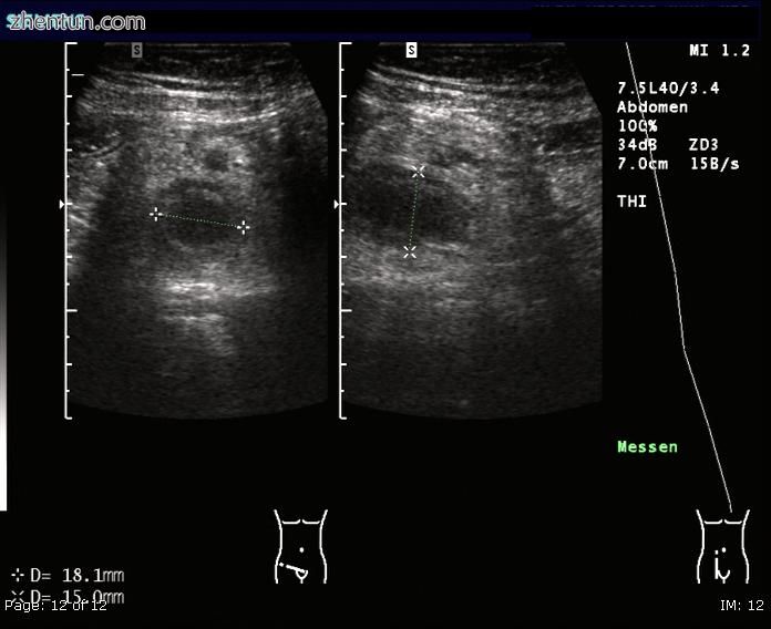
超声图像急性阑尾炎
腹部超声检查,优选用多普勒超声检查,可用于检测阑尾炎,特别是在儿童中。在使用彩色多普勒时,超声可以显示右侧髂窝内游离液体收集情况,以及可见的阑尾血流量增加和阑尾不可压缩性,因为它本质上是壁脓肿。急性阑尾炎的其他继发超声征象包括阑尾周围的回声性肠系膜脂肪和阑尾的声影。[46]在某些情况下(约5%)[47],尽管存在阑尾炎,髂窝超声检查仍未发现任何异常。在阑尾明显扩张之前,这种假阴性结果尤其适用于早期阑尾炎。此外,在大量脂肪和肠道气体使附件在技术上难以可视化的成人中,假阴性结果更常见。尽管有这些限制,但有经验的手中的超声成像通常可区分阑尾炎和其他类似症状的疾病。其中一些病症包括阑尾附近淋巴结的炎症或来源于其他盆腔器官(如卵巢或输卵管)的疼痛。
文件:UOTW 45 - 本周超声1.webm
https://cache.tv.qq.com/qqplayerout.swf?vid=f0705zqvuha
超声显示阑尾炎和阑尾[48]
Ultrasound showing appendicitis and an appendicolith[48]
![Ultrasound showing appendicitis and an appendicolith[48] Ultrasound showing appendicitis and an appendicolith[48]](data/attachment/forum/201806/28/160058pt26g11r6ewge81f.jpg)
超声显示阑尾炎和阑尾[48]
Ultrasound of a normal appendix for comparison
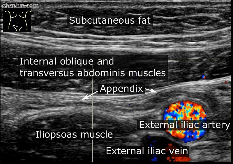
正常阑尾的超声波比较
A normal appendix without and with compression. Absence of comprehensibility indicates appendicitis. ...
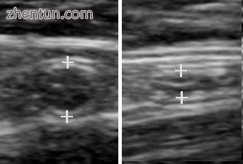
没有压缩的正常附件。 缺乏可理解性表明阑尾炎。[46]
CT检查
A CT scan demonstrating acute appendicitis (note the appendix has a diameter of 17.1 mm and there is ...
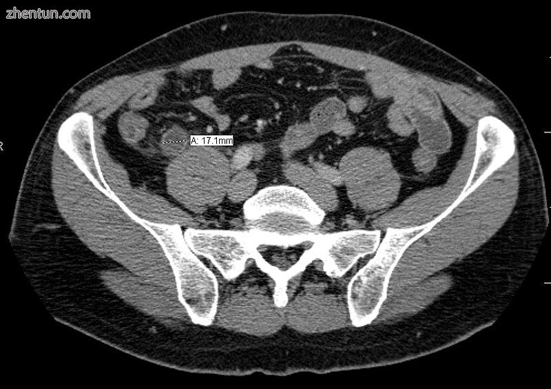
CT扫描显示急性阑尾炎(注意阑尾直径为17.1mm,周围有脂肪堆积)
A fecalith marked by the arrow that has resulted in acute appendicitis.
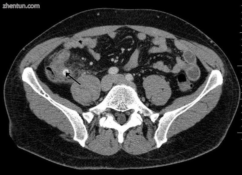
由箭头标记的粪便导致急性阑尾炎。
计算机断层扫描(CT)容易获得,特别是对于那些诊断历史和体格检查不明显的人来说,它已经被广泛使用。对辐射的担忧往往限制孕妇和儿童使用CT,特别是随着MRI的使用日益广泛[49] [50]。
阑尾炎的准确诊断是多层次的,阑尾的大小具有最强的阳性预测值,而间接特征可以增加或降低敏感性和特异性。超过6毫米的尺寸对阑尾炎敏感和特异性都很高[51]。
然而,因为阑尾可以充满粪便,引起腔内扩张,所以这个标准在更近期的荟萃分析中显示出有限的效用[52]。这与超声相反,其中阑尾的壁可以更容易地与腔内粪便区分。在这种情况下,辅助功能如与邻近肠道相比增加了壁增强和周围脂肪的炎症或脂肪搁浅可以支持诊断,尽管他们的缺席并不排除它。在穿孔严重的情况下,可以看到相邻的蜂窝织炎或脓肿。也可能导致骨盆中稠密的液体分层,与脓液或肠道溢出有关。当病人瘦小或年轻时,相对缺乏脂肪会使阑尾和周围的脂肪徘徊难以看清[52]。
磁共振成像
由于辐射剂量,MRI对儿童和怀孕患者阑尾炎的诊断越来越普遍,尽管健康成年人的风险几乎可以忽略不计,但可能对儿童或发育中的胎儿有害。在怀孕期间,发现它在妊娠中期和晚期更有用,特别是当扩大的子宫取代了阑尾时,很难通过超声发现。 MRI上脂肪绞合导致的CT上反映的阑尾周围的绞合表现为T2加权序列上流体信号增加。孕早期妊娠通常不是MRI的候选对象,因为胎儿仍在进行器官发生,至今尚未对其潜在风险或副作用进行长期研究[53]。
X线
Appendicolith as seen on plain X-ray
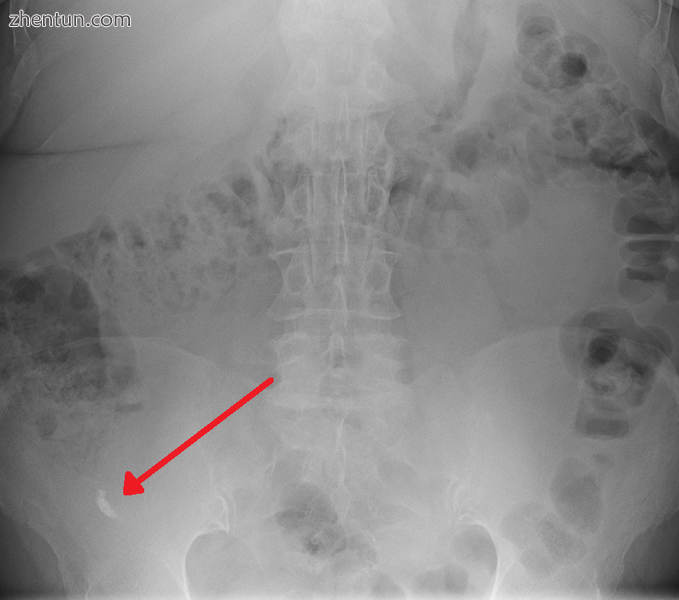
平片可见的阑尾结石
一般来说,腹部平片造影术(PAR)对阑尾炎的诊断并不有用,也不应该从接受阑尾炎评估的人那里常规获得[54] [55]。单纯的腹部薄膜可用于检测输尿管结石,小肠梗阻或穿孔性溃疡,但这些情况很少与阑尾炎相混淆[56]。在不到5%的被评估为阑尾炎的人群中,在右下腹可以确定一种不透明的粪便[40]。钡剂灌肠已被证明是阑尾炎的一种较差的诊断工具。虽然钡剂灌肠时阑尾填塞失败与阑尾炎有关,但正常附件的20%不能填充[56]。
评分系统
没有出色的评分系统来确定一个孩子是否患有阑尾炎[57]。 Alvarado评分和儿科阑尾炎评分是有用的,但并不是确定性的[57]。
Alvarado评分是最广泛使用的评分系统。低于5的分数表明对阑尾炎的诊断,而7分或更高的分数可预测急性阑尾炎。对于可疑得分为5或6的患者,可以使用CT扫描或超声检查来降低阴性阑尾切除术的发生率。
| Alvarado评分 | | | 迁徙性右髂窝疼痛 | 1分 | | 厌食 | 1分 | | 恶心和呕吐 | 1分 | | 右髂窝压痛 | 2分 | | 反跳性腹部压痛 | 1分 | | 发热 | 1分 | | 高白细胞计数(白细胞增多症) | 2分 | | 向左移(分割的嗜中性粒细胞) | 1分 | | 总得分 | 10分 |
明确的诊断是基于病理学的。 阑尾炎的组织学发现是固有肌层的中性粒细胞浸润。
阑尾周围炎,阑尾周围组织的炎症常常与其他腹部病理学一起发现[58]。
Micrograph of appendicitis and periappendicitis. H&E stain.
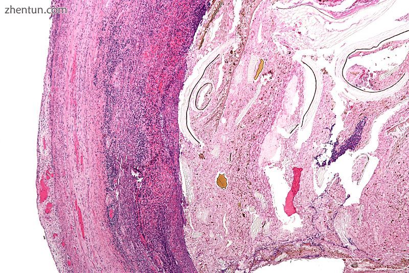
阑尾炎和阑尾炎的显微照片。H&E染色。
Micrograph of appendicitis showing neutrophils in the muscularis propria. H&E stain.
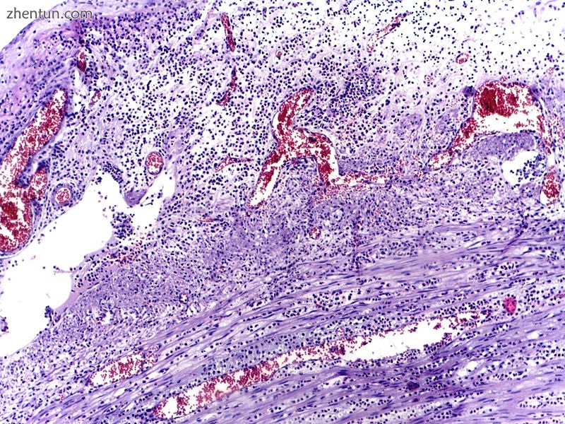
阑尾炎显微照片显示固有肌层中性粒细胞。H&E染色。
鉴别诊断
Coronal CT scan of a person initially suspected of having appendicitis because of right-sided pain. ...
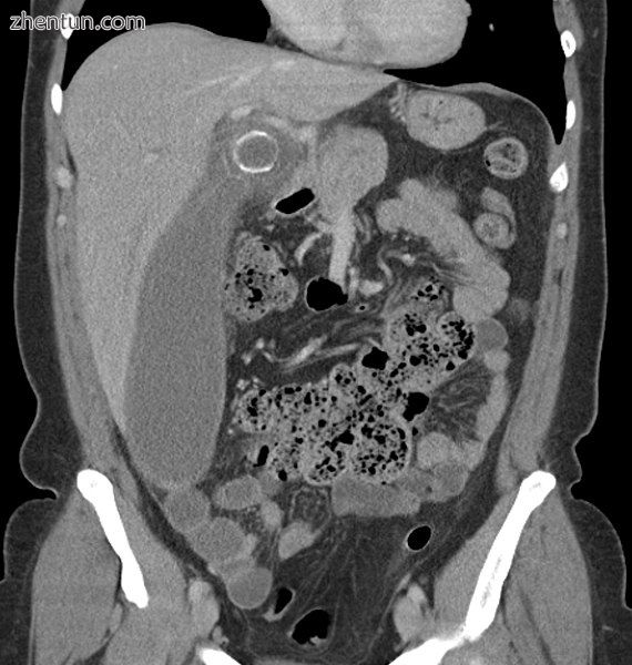
由于右侧疼痛,最初怀疑患有阑尾炎的人的冠状CT扫描。 CT显示实际上是一个扩大的发炎胆囊,到达右下腹部。
儿童:胃肠炎,肠系膜腺炎,梅克尔憩室炎,肠套叠,过敏性紫癜,大叶性肺炎,尿路感染(UTI患儿可出现其他症状时无腹痛),新发克罗恩病或溃疡性结肠炎,胰腺炎,虐待儿童的腹部创伤;囊性纤维化患儿远端肠梗阻综合征;儿童白血病的危害。
女性:妊娠试验对所有育龄妇女都很重要,因为宫外孕的症状和体征与阑尾炎相似。其他妇产科/妇科病因包括盆腔炎,卵巢扭转,月经初潮,痛经,子宫内膜异位症和Mittelschmerz(大约在月经前两周卵子通过)[59]。
男性:睾丸扭转
成人:新发克隆氏病,溃疡性结肠炎,局部肠炎,胆囊炎,肾绞痛,穿孔消化性溃疡,胰腺炎,直肠鞘膜血肿和外翻性附件炎。
老年人:憩室炎,肠梗阻,结肠癌,肠系膜缺血,漏主动脉瘤。
术语“假性阑尾炎”用于描述模仿阑尾炎的病症。[60]它可以与小肠结肠炎耶尔森氏菌相关[61]。
管理
急性阑尾炎通常由手术管理。虽然抗生素治疗无并发症的阑尾炎是安全和有效的,[7] [62] 26%的人一年内复发并需要最终的阑尾切除术[63]。如果有阑尾存在,抗生素疗效就会降低[64]。手术是急性阑尾炎的标准治疗方法[65]。手术与抗生素的成本效益尚不清楚[66]。
建议使用抗生素预防急诊阑尾切除手术中潜在的术后并发症,并且在手术前,手术过程中或手术后给予人抗生素是有效的[67]。
疼痛
疼痛药物(如吗啡)似乎不会影响临床诊断阑尾炎的准确性,因此应该在患者的护理早期给予。历史上,一些普通外科医生担心镇痛剂会影响儿童的临床检查,有些医生建议直到外科医生能够检查患者时才能给予止痛药。
Inflamed appendix removal by open surgery
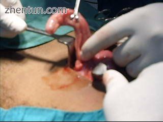
开放手术切除炎症阑尾
Laparoscopic appendectomy.
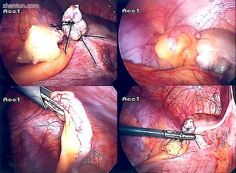
腹腔镜阑尾切除术。
去除阑尾的手术过程称为阑尾切除术。阑尾切除术可以通过开放式或腹腔镜手术进行。腹腔镜阑尾切除术作为急性阑尾炎的介入治疗与开腹阑尾切除术相比有几个优点。
开放阑尾切除术
一个多世纪以来,开腹手术(开放式阑尾切除术)是急性阑尾炎的标准治疗方法[70]。该过程包括通过腹部右下区域的单个大切口去除感染的阑尾[71]。剖腹手术中的切口通常长2至3英寸(51至76毫米)。
在开腹阑尾切除术期间,疑似阑尾炎的患者置于全身麻醉下,以保持肌肉完全放松并保持该人无意识。切口长约2至3英寸(76毫米),并在右下腹部形成,在臀骨上方数英寸处。一旦切口打开腹腔并确定阑尾,外科医生将被感染的组织切除并从附近的组织切下阑尾。仔细检查感染区后,确保没有迹象表明周围组织受到损伤或感染。如果由急诊开放性阑尾切除术治疗复杂性阑尾炎,可以插入腹部引流(从腹部到外部的临时管以避免脓肿形成),但这可能会增加住院时间[72]。外科医生将开始关闭切口。这意味着缝合肌肉并使用手术缝合钉或针缝合皮肤。为了防止感染,切口覆盖无菌绷带。
腹腔镜阑尾切除术
腹腔镜阑尾切除术自1983年引入以来,已成为急性阑尾炎日益普遍的干预措施。[73]这种外科手术包括在腹部做三至四个切口,每个切口长0.25至0.5英寸(6.4至12.7毫米)。这种类型的阑尾切除术是通过将一种称为腹腔镜的特殊手术工具插入其中一个切口中而制成的。腹腔镜连接到人体外的显示器,旨在帮助外科医生检查腹部的感染区域。另外两个切口用于通过使用手术器械来特定移除附件。腹腔镜手术需要全身麻醉,并且可以持续两个小时。腹腔镜阑尾切除术与开放式阑尾切除术相比有几个优点,包括手术后恢复时间短,术后疼痛少,浅表手术部位感染率低。然而,在腹腔镜阑尾切除术中腹腔脓肿的发生率几乎是开腹阑尾切除术的三倍[74]。
手术前
治疗始于将患有手术的人禁食或饮食一段时间,通常是过夜。静脉滴注用于为将进行手术的人提供水分。静脉给予抗生素,如头孢呋辛和甲硝唑可以早期给药,以帮助杀死细菌,从而减少感染在腹部的扩散以及腹部或伤口的术后并发症。模棱两可的病例可能变得更难以用抗生素治疗进行评估,并从系列检查中受益。如果胃空着(过去六小时内没有食物),通常使用全身麻醉。否则,可以使用脊髓麻醉。
一旦决定进行阑尾切除术,准备程序大约需要一到两个小时。同时,外科医生将解释手术过程,并将介绍执行阑尾切除术时必须考虑的风险。 (对于所有手术,在执行手术之前都存在必须评估的风险。)风险根据附录的状态而有所不同。如果阑尾未破裂,并发症发生率仅为3%左右,但如果阑尾破裂,并发症发生率将上升至近59%[75]。可能发生的最常见的并发症是肺炎,切口疝,血栓性静脉炎,出血或粘连。最近的证据表明,入院后延迟获得手术结果与阑尾炎患者的结局没有明显的差异[76] [77]。
外科医生将解释恢复过程需要多长时间。通常会移除腹部的头发以避免可能出现的关于切口的并发症。
在大多数情况下,进行手术的患者在手术前会遇到需要用药的恶心或呕吐。在阑尾切除术之前可以使用抗生素和止痛药物。
手术后
The stitches the day after having the appendix removed by laparoscopic surgery
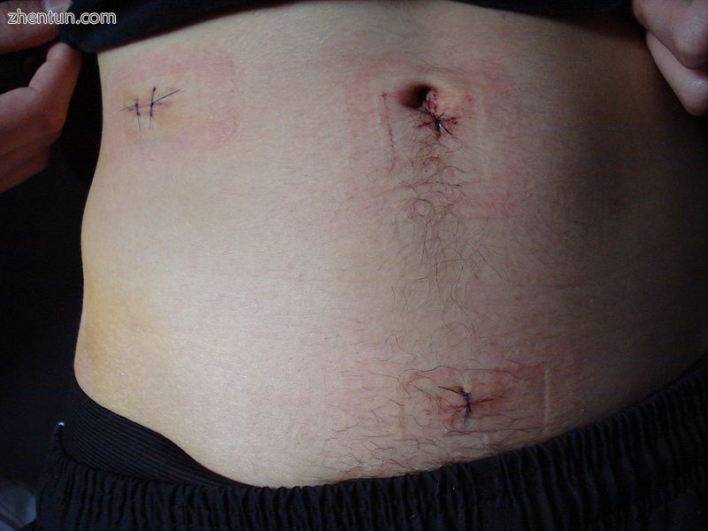
腹腔镜手术切除阑尾后第二天缝合
住院时间通常为几小时至几天,但如果出现并发症,可能需要几周时间。根据病情的严重程度,康复过程可能会有所不同:手术前阑尾是否破裂。如果阑尾未破裂,附录手术恢复一般要快得多。接受手术的人尊重医生的建议并限制他们的体力活动,这样组织可以愈合得更快,这一点很重要。阑尾切除术后恢复可能不需要改变饮食习惯或改变生活方式。
阑尾炎住院时间因病情的严重程度而异。来自美国的一项研究发现,2010年,平均阑尾炎住院时间为1.8天。对于阑尾破裂的住院病人,平均住院时间为5.2天[13]。
手术后,病人将被转移到麻醉后监护病房,以便密切监测他或她的生命体征,以检测麻醉或手术相关并发症。必要时可以服用止痛药。患者完全清醒后,他们被转移到医院房间恢复。大多数人在手术后第二天都会提供清澈的液体,然后在肠子开始正常运转时进行正常饮食。建议患者坐在床边,每天短距离走几次。移动是强制性的,必要时可给予止痛药。完全从阑尾切除术恢复大约需要四到六周,但如果阑尾破裂,可以延长至八周。
预测
大多数阑尾炎患者在手术治疗后很容易恢复,但如果延误治疗或发生腹膜炎,可能会出现并发症。恢复时间取决于年龄,病情,并发症和其他情况,包括饮酒量,但通常在10到28天之间。对于年幼的儿童(大约10岁),恢复需要三周的时间。
腹膜炎的可能性是急性阑尾炎需要快速评估和治疗的原因。疑似阑尾炎的人可能需要接受医疗后送。有时在紧急情况下(即不在适当的医院)进行阑尾切除术,因为不能及时进行医疗撤离。
典型的急性阑尾炎迅速对阑尾切除术作出反应,偶尔会自发消退。如果阑尾炎自发消退,是否应该进行选择性间期阑尾切除术以预防阑尾炎反复发作仍存在争议。非典型阑尾炎(与化脓性阑尾炎相关)更难以诊断,即使在早期手术时也更容易变得复杂。在任何一种情况下,即刻诊断和阑尾切除术产生最佳结果,通常在2至4周内完全康复。死亡率和严重并发症是罕见的,但确实发生,特别是如果腹膜炎持续存在并且未被治疗。
被称为阑尾肿块的另一个实体被讨论了。 它发生在感染期间阑尾未被清除并且大网膜和肠粘连时,形成可触及的肿块。 在此期间,手术是有风险的,除非发热和毒性或USG导致脓液形成。 医疗管理对待病症。
阑尾切除术的一个不寻常的并发症是“残端阑尾炎”:炎症发生在先前不完全阑尾切除术后留下的残留阑尾残端。 阑尾炎可能会在阑尾切除术后数月至数年发生,并且可以通过超声等成像方式进行鉴定[80]。
流行病学
Appendicitis deaths per million persons in 2012
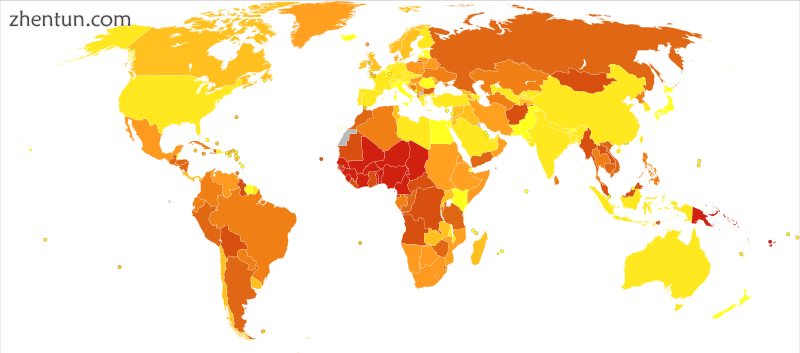
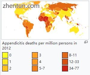
2012年每百万人阑尾炎死亡人数
Disability-adjusted life year for appendicitis per 100,000 inhabitants in 2004.[81]
![Disability-adjusted life year for appendicitis per 100,000 inhabitants in 2004.[81] Disability-adjusted life year for appendicitis per 100,000 inhabitants in 2004.[81]](data/attachment/forum/201806/28/160044hq0spc2c27icluqd.png)
2004年每10万居民阑尾炎的残疾调整生命年。[81]
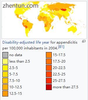
阑尾炎在5至40岁之间最常见; [82]中间年龄为28岁。危险因素包括男性,家庭收入较高以及生活在农村地区[83]。 2013年,全球死亡人数从1990年的88,000人下降至全球72,000人。[84]
参考:
1. "appendicitis". Medical Dictionary. Merriam-Webster. Archived from the original on 2013-12-30.
2. Graffeo, Charles S.; Counselman, Francis L. (November 1996). "Appendicitis". Emergency Medicine Clinics of North America. 14 (4): 653–71. doi:10.1016/s0733-8627(05)70273-x. PMID 8921763.
3. Hobler, K. (Spring 1998). "Acute and Suppurative Appendicitis: Disease Duration and its Implications for Quality Improvement" (PDF). Permanente Medical Journal. 2.
4. Paulson, EK; Kalady, MF; Pappas, TN (16 January 2003). "Clinical practice. Suspected appendicitis" (PDF). The New England Journal of Medicine. 348 (3): 236–42. doi:10.1056/nejmcp013351. PMID 12529465.
5. Ferri, Fred F. (2010). Ferri's differential diagnosis : a practical guide to the differential diagnosis of symptoms, signs, and clinical disorders (2nd ed.). Philadelphia, PA: Elsevier/Mosby. p. Chapter A. ISBN 0323076998.
6. Longo, Dan L.; et al., eds. (2012). Harrison's principles of internal medicine (18th ed.). New York: McGraw-Hill. p. Chapter 300. ISBN 978-0-07174889-6. Archived from the original on 30 March 2016. Retrieved 6 November 2014.
7. Varadhan KK, Neal KR, Lobo DN (2012). "Safety and efficacy of antibiotics compared with appendicectomy for treatment of uncomplicated acute appendicitis: meta-analysis of randomised controlled trials". The BMJ. 344: e2156. doi:10.1136/bmj.e2156. PMC 3320713 Freely accessible. PMID 22491789.
8. GBD 2015 Disease and Injury Incidence and Prevalence, Collaborators. (8 October 2016). "Global, regional, and national incidence, prevalence, and years lived with disability for 310 diseases and injuries, 1990-2015: a systematic analysis for the Global Burden of Disease Study 2015". Lancet. 388 (10053): 1545–1602. doi:10.1016/S0140-6736(16)31678-6. PMC 5055577 Freely accessible. PMID 27733282.
9. GBD 2015 Mortality and Causes of Death, Collaborators. (8 October 2016). "Global, regional, and national life expectancy, all-cause mortality, and cause-specific mortality for 249 causes of death, 1980-2015: a systematic analysis for the Global Burden of Disease Study 2015". Lancet. 388 (10053): 1459–1544. doi:10.1016/s0140-6736(16)31012-1. PMC 5388903 Freely accessible. PMID 27733281.
10. Pieper R, Kager L, Tidefeldt U (1982). "Obstruction of appendix vermiformis causing acute appendicitis. One of the most common causes of this is an acute viral infection which causes lymphoid hyperplasia and therefore obstruction. An experimental study in the rabbit". Acta Chirurgica Scandinavica. 148 (1): 63–72. PMID 7136413.
11. Tintinalli, editor-in-chief Judith E. (2011). Emergency medicine : a comprehensive study guide (7. ed.). New York: McGraw-Hill. p. Chapter 84. ISBN 978-0-07-174467-6. Archived from the original on 22 December 2016. Retrieved 6 November 2014.
12. Schwartz's principles of surgery (9th ed.). New York: McGraw-Hill, Medical Pub. Division. 2010. p. Chapter 30. ISBN 978-0-07-1547703.
13. Barrett, ML; Hines, AL; Andrews, RM (July 2013). "Trends in Rates of Perforated Appendix, 2001–2010" (PDF). Healthcare Cost and Utilization Project Statistical Brief #159. Agency for Healthcare Research and Quality. PMID 24199256. Archived (PDF) from the original on 2016-10-20.
14. Shogilev, DJ; Duus, N; Odom, SR; Shapiro, NI (November 2014). "Diagnosing appendicitis: evidence-based review of the diagnostic approach in 2014". The Western Journal of Emergency Medicine (Review). 15 (7): 859–71. doi:10.5811/westjem.2014.9.21568. PMC 4251237 Freely accessible. PMID 25493136.
15. Mason, RJ (August 2008). "Surgery for appendicitis: is it necessary?". Surgical Infections. 9 (4): 481–8. doi:10.1089/sur.2007.079. PMID 18687030.
16. Fitz RH (1886). "Perforating inflammation of the vermiform appendix with special reference to its early diagnosis and treatment". American Journal of Medical Science (92): 321–46.
17. Wangensteen OH, Bowers WF (1937). "Significance of the obstructive factor in the genesis of acute appendicitis". Archives of Surgery. 34 (3): 496–526. doi:10.1001/archsurg.1937.01190090121006.
18. Hollerman, J.; Bernstein, MA; Kottamasu, SR; Sirr, SA (1988). "Acute recurrent appendicitis with appendicolith". The American Journal of Emergency Medicine. 6 (6): 614–617. doi:10.1016/0735-6757(88)90105-2.
19. Dehghan A, Moaddab AH, Mozafarpour S (June 2011). "An unusual localization of trichobezoar in the appendix". The Turkish Journal of Gastroenterology. 22 (3): 357–8. PMID 21805435.
20. Jones BA, Demetriades D, Segal I, Burkitt DP (1985). "The prevalence of appendiceal fecaliths in patients with and without appendicitis. A comparative study from Canada and South Africa". Annals of Surgery. 202 (1): 80–82. doi:10.1097/00000658-198507000-00013. PMC 1250841 Freely accessible. PMID 2990360.
21. Nitecki S, Karmeli R, Sarr MG (1990). "Appendiceal calculi and fecaliths as indications for appendectomy". Surgery, Gynecology & Obstetrics. 171 (3): 185–8. PMID 2385810.
22. Arnbjörnsson E (1985). "Acute appendicitis related to faecal stasis". Annales Chirurgiae et Gynaecologiae. 74 (2): 90–3. PMID 2992354.
23. Raahave D, Christensen E, Moeller H, Kirkeby LT, Loud FB, Knudsen LL (2007). "Origin of acute appendicitis: fecal retention in colonic reservoirs: a case control study". Surgical Infections. 8 (1): 55–62. doi:10.1089/sur.2005.04250. PMID 17381397.
24. Burkitt DP (1971). "The aetiology of appendicitis". British Journal of Surgery. 58 (9): 695–9. doi:10.1002/bjs.1800580916. PMC 1598350 Freely accessible. PMID 4937032.
25. Segal I, Walker AR (1982). "Diverticular disease in urban Africans in South Africa". Digestion. 24 (1): 42–6. doi:10.1159/000198773. PMID 6813167.
26. Arnbjörnsson E (1982). "Acute appendicitis as a sign of a colorectal carcinoma". Journal of Surgical Oncology. 20 (1): 17–20. doi:10.1002/jso.2930200105. PMID 7078180.
27. Burkitt DP, Walker AR, Painter NS (1972). "Effect of dietary fibre on stools and the transit-times, and its role in the causation of disease". The Lancet. 2 (7792): 1408–12. doi:10.1016/S0140-6736(72)92974-1. PMID 4118696.
28. Adamis D, Roma-Giannikou E, Karamolegou K (2000). "Fiber intake and childhood appendicitis". International Journal of Food Science and Nutrition. 51 (3): 153–7. doi:10.1080/09637480050029647. PMID 10945110.
29. Hugh TB, Hugh TJ (2001). "Appendicectomy--becoming a rare event?". Medical Journal of Australia. 175 (1): 7–8. PMID 11476215. Archived from the original on 2006-08-26.
30. Gear JS, Brodribb AJ, Ware A, Mann JI (January 1981). "Fibre and bowel transit times". The British Journal of Nutrition. 45 (1): 77–82. doi:10.1079/BJN19810078. PMID 6258626.
31. Ashdown, H. F.; D'Souza, N.; Karim, D.; Stevens, R. J.; Huang, A.; Harnden, A. (17 December 2012). "Pain over speed bumps in diagnosis of acute appendicitis: diagnostic accuracy study". The BMJ. 345 (dec14 14): e8012–e8012. doi:10.1136/bmj.e8012. PMC 3524367 Freely accessible. PMID 23247977.
32. Hobler, K. (Spring 1998). "Acute and Suppurative Appendicitis: Disease Duration and its Implications for Quality Improvement" (PDF). Permanente Medical Journal. 2 (2). Archived from the original (PDF) on 2008-10-07.
33. Sachdeva, Anupam; Dutta, AK (31 August 2012). Advances in Pediatrics. JP Medical. p. 1432. ISBN 9789350257777.
34. Small, V (2008). Dolan, B; Holt, L, eds. Surgical emergencies. Accident and Emergency: Theory into Practice (2nd ed.). Elsevier.[page needed]
35. Virgilio, Christian de; Frank, Paul N.; Grigorian, Areg (10 January 2015). Surgery. Springer. p. 215. ISBN 9781493917266.
36. Kadim, Abbas Abdul Mahdi (1 April 2016). "Surgical and Clinical Review of Acute Appendicitis" (PDF). International Journal of Multidisciplinary and Current Research. 4. ISSN 2321-3124. Archived (PDF) from the original on 9 August 2016. Massouh sign characterized by increased abdominal pain with coughing. It may be an indicator of appendicitis. A positive Massouh sign is a grimace of the person being examined upon a right sided (and not left) sweep (40).
37. Wolfson, Allan B.; Cloutier, Robert L.; Hendey, Gregory W.; Ling, Louis J.; Schaider, Jeffrey J.; Rosen, Carlo L. (27 October 2014). Harwood-Nuss' Clinical Practice of Emergency Medicine. Wolters Kluwer Health. p. 5810. ISBN 9781469889481. Archived from the original on 10 September 2017. Retrieved 15 June 2016. Physical signs classically associated with acute appendicitis include Rovsing sign, psoas sign, and obturator sign.
38. Rovsing, N.T. (1907). "Indirektes Hervorrufen des typischen Schmerzes an McBurney's Punkt. Ein Beitrag zur diagnostik der Appendicitis und Typhlitis". Zentralblatt für Chirurgie. Leipzig. 34: 1257–1259.(in German)
39. Dunster, Edward Swift; Hunter, James Bradbridge; Sajous, Charles Euchariste de Medicis; Foster, Frank Pierce; Stragnell, Gregory; Klaunberg, Henry J.; Martí-Ibáñez, Félix (1922). International Record of Medicine and General Practice Clinics. New York Medical Journal. p. 663.
40. Gregory, Charmaine (2010). "Appendicitis". CDEM Self Study Modules. Clerkship Directors in Emergency Medicine. Archived from the original on 2013-11-30.
41. Bundy DG, Byerley JS, Liles EA, Perrin EM, Katznelson J, Rice HE (2007). "Does this child have appendicitis?". JAMA. 298 (4): 438–51. doi:10.1001/jama.298.4.438. PMC 2703737 Freely accessible. PMID 17652298.
42. American College of Radiology, "Five Things Physicians and Patients Should Question" (PDF), Choosing Wisely: an initiative of the ABIM Foundation, American College of Radiology, archived (PDF) from the original on April 16, 2012, retrieved August 17, 2012
43. Krishnamoorthi, R.; Ramarajan, N.; Wang, N. E.; Newman, B.; Rubesova, E.; Mueller, C. M.; Barth, R. A. (2011). "Effectiveness of a Staged US and CT Protocol for the Diagnosis of Pediatric Appendicitis: Reducing Radiation Exposure in the Age of ALARA". Radiology. 259 (1): 231–239. doi:10.1148/radiol.10100984. PMID 21324843.
44. Wan, M. J.; Krahn, M.; Ungar, W. J.; Caku, E.; Sung, L.; Medina, L. S.; Doria, A. S. (2009). "Acute Appendicitis in Young Children: Cost-effectiveness of US versus CT in Diagnosis--A Markov Decision Analytic Model". Radiology. 250 (2): 378–386. doi:10.1148/radiol.2502080100. PMID 19098225.
45. Terasawa T, Blackmore CC, Bent S, Kohlwes RJ (October 2004). "Systematic review: computed tomography and ultrasonography to detect acute appendicitis in adults and adolescents". Annals of Internal Medicine. 141 (7): 537–46. doi:10.7326/0003-4819-141-7-200410050-00011. PMID 15466771.
46. Reddan, Tristan; Corness, Jonathan; Mengersen, Kerrie; Harden, Fiona (March 2016). "Ultrasound of paediatric appendicitis and its secondary sonographic signs: providing a more meaningful finding". Journal of Medical Radiation Sciences. 63 (1): 59–66. doi:10.1002/jmrs.154.
47. Reddan, Tristan; Corness, Jonathan; Mengersen, Kerrie; Harden, Fiona (June 2016). "Sonographic diagnosis of acute appendicitis in children: a 3-year retrospective". Sonography. doi:10.1002/sono.12068.
48. "UOTW #45 - Ultrasound of the Week". Ultrasound of the Week. 25 April 2015. Archived from the original on 9 May 2017.
49. Kim, Y; Kang, G; Moon, SB (November 2014). "Increasing utilization of abdominal CT in the Emergency Department of a secondary care center: does it produce better outcomes in caring for pediatric surgical patients?". Annals of Surgical Treatment and Research. 87 (5): 239–44. doi:10.4174/astr.2014.87.5.239. PMC 4217253 Freely accessible. PMID 25368849.
50. Liu, B; Ramalho, M; AlObaidy, M; Busireddy, KK; Altun, E; Kalubowila, J; Semelka, RC (28 August 2014). "Gastrointestinal imaging-practical magnetic resonance imaging approach". World Journal of Radiology. 6 (8): 544–66. doi:10.4329/wjr.v6.i8.544. PMC 4147436 Freely accessible. PMID 25170393.
51. Garcia, K.; Hernanz-Schulman, M.; Bennett, D. L.; Morrow, S. E.; Yu, C.; Kan, J. H. (2009). "Suspected Appendicitis in Children: Diagnostic Importance of Normal Abdominopelvic CT Findings with Nonvisualized Appendix". Radiology. 250 (2): 531–537. doi:10.1148/radiol.2502080624. PMID 19188320.
52. Doria, A. S.; Moineddin, R.; Kellenberger, C. J.; Epelman, M.; Beyene, J.; Schuh, S.; Babyn, P. S.; Dick, P. T. (2006). "US or CT for Diagnosis of Appendicitis in Children and Adults? A Meta-Analysis". Radiology. 241 (1): 83–94. doi:10.1148/radiol.2411050913. PMID 16928974.
53. Burke, LM; Bashir, MR; Miller, FH; Siegelman, ES; Brown, M; Alobaidy, M; Jaffe, TA; Hussain, SM; Palmer, SL; Garon, BT; Oto, A; Reinhold, C; Ascher, SM; Demulder, DK; Thomas, S; Best, S; Borer, J; Zhao, K; Pinel-Giroux, F; De Oliveira, I; Resende, D; Semelka, RC (24 July 2015). "Magnetic Resonance Imaging of Acute Appendicitis in Pregnancy: a 5-Year Multi-institutional Study". American Journal of Obstetrics and Gynecology. 213: 693.e1–6. doi:10.1016/j.ajog.2015.07.026. PMID 26215327.
54. Rao PM, Rhea JT, Rao JA, Conn AK (July 1999). "Plain abdominal radiography in clinically suspected appendicitis: diagnostic yield, resource use, and comparison with CT". The American Journal of Emergency Medicine. 17 (4): 325–8. doi:10.1016/S0735-6757(99)90077-3. PMID 10452424.
55. Boleslawski E, Panis Y, Benoist S, Denet C, Mariani P, Valleur P (March 1999). "Plain abdominal radiography as a routine procedure for acute abdominal pain of the right lower quadrant: prospective evaluation". World Journal of Surgery. 23 (3): 262–4. doi:10.1007/pl00013181. PMID 9933697.
56. APPENDICITIS from Townsend: Sabiston Textbook of Surgery on MD Consult Archived December 3, 2013, at the Wayback Machine.
57. Kulik, DM; Uleryk, EM; Maguire, JL (January 2013). "Does this child have appendicitis? A systematic review of clinical prediction rules for children with acute abdominal pain". Journal of Clinical Epidemiology. 66 (1): 95–104. doi:10.1016/j.jclinepi.2012.09.004. PMID 23177898.
58. Fink AS, Kosakowski CA, Hiatt JR, Cochran AJ (June 1990). "Periappendicitis is a significant clinical finding". American Journal of Surgery. 159 (6): 564–8. doi:10.1016/S0002-9610(06)80067-X. PMID 2349982.
59. "Pelvic inflammatory disease (PID) Symptoms; Diseases and Conditions". Mayo Clinic. Archived from the original on 2015-05-07. Retrieved 2015-04-23.
60. Cunha BA, Pherez FM, Durie N (July 2010). "Swine influenza (H1N1) and acute appendicitis". Heart Lung. 39 (6): 544–6. doi:10.1016/j.hrtlng.2010.04.004. PMID 20633930.
61. Zheng H, Sun Y, Lin S, Mao Z, Jiang B (August 2008). "Yersinia enterocolitica infection in diarrheal patients". Eur. J. Clin. Microbiol. Infect. Dis. 27 (8): 741–52. doi:10.1007/s10096-008-0562-y. ISBN 0-9600805-6-2. PMID 18575909.
62. Sallinen, V; Akl, EA; You, JJ; Agarwal, A; Shoucair, S; Vandvik, PO; Agoritsas, T; Heels-Ansdell, D; Guyatt, GH; Tikkinen, KA (17 March 2016). "Meta-analysis of antibiotics versus appendicectomy for non-perforated acute appendicitis". The British Journal of Surgery. 103: 656–667. doi:10.1002/bjs.10147. PMID 26990957.
63. Harnoss, JC; Zelienka, I; Probst, P; Grummich, K; Müller-Lantzsch, C; Harnoss, JM; Ulrich, A; Büchler, MW; Diener, MK (17 October 2016). "Antibiotics Versus Surgical Therapy for Uncomplicated Appendicitis: Systematic Review and Meta-analysis of Controlled Trials (PROSPERO 2015: CRD42015016882)". Annals of Surgery: 1. doi:10.1097/SLA.0000000000002039. PMID 27759621.
64. Huang, Libin; Yin, Yuan; Yang, Lie; Wang, Cun; Li, Yuan; Zhou, Zongguang (27 March 2017). "Comparison of Antibiotic Therapy and Appendectomy for Acute Uncomplicated Appendicitis in Children". JAMA Pediatrics. doi:10.1001/jamapediatrics.2017.0057.
65. Wilms, IM; de Hoog, DE; de Visser, DC; Janzing, HM (9 November 2011). "Appendectomy versus antibiotic treatment for acute appendicitis". The Cochrane Database of Systematic Reviews (11): CD008359. doi:10.1002/14651858.CD008359.pub2. PMID 22071846.
66. Georgiou, R; Eaton, S; Stanton, MP; Pierro, A; Hall, NJ (March 2017). "Efficacy and Safety of Nonoperative Treatment for Acute Appendicitis: A Meta-analysis". Pediatrics. 139 (3): e20163003. doi:10.1542/peds.2016-3003. PMID 28213607.
67. Andersen, BR; Kallehave, FL; Andersen, HK (20 July 2005). "Antibiotics versus placebo for prevention of postoperative infection after appendicectomy". The Cochrane Database of Systematic Reviews (3): CD001439. doi:10.1002/14651858.CD001439.pub2. PMID 16034862.
68. Anderson, Mark; Collins, Emma (November 2008). "Analgesia for children with acute abdominal pain and diagnostic accuracy". Archives of Disease in Childhood. 93 (11): 995–7. doi:10.1136/adc.2008.137174. PMID 18305071. Archived from the original on 2013-05-17.
69. Sauerland, S; Jaschinski, T; Neugebauer, EA (Oct 6, 2010). "Laparoscopic versus open surgery for suspected appendicitis". Cochrane Database of Systematic Reviews (10): CD001546. doi:10.1002/14651858.CD001546.pub3. PMID 20927725.
70. Berry J, Malt RA (1984). "Appendicitis near its centenary". Annals of Surgery. 200: 567–75. doi:10.1097/00000658-198411000-00002. PMC 1250537 Freely accessible. PMID 6385879. More than one of |pmc= and |PMC= specified (help)
71. "Appendicitis". National Institute of Diabetes and Digestive and Kidney Diseases. U.S Department of Health and Human Services. Archived from the original on 2010-02-01. Retrieved 2010-02-01.
72. Cheng, Y; Zhou, S; Zhou, R; Lu, J; Wu, S; Xiong, X; Ye, H; Lin, Y; Wu, T; Cheng, N (7 February 2015). "Abdominal drainage to prevent intra-peritoneal abscess after open appendectomy for complicated appendicitis". The Cochrane Database of Systematic Reviews (2): CD010168. doi:10.1002/14651858.CD010168.pub2. PMID 25914903.
73. Semm K (March 1983). "Endoscopic appendectomy". Endoscopy. 15: 59–64. doi:10.1055/s-2007-1021466. PMID 6221925.
74. Siewert B, Raptopoulos V, Liu SI, Hodin RA, Davis RB, Rosen MP (2003). "CT predictors of failed laparoscopic appendectomy". Radiology. 229: 415–20. doi:10.1148/radiol.2292020825. PMID 14595145.
75. "Appendicitis". Encyclopedia of Surgery. Archived from the original on 2010-02-09. Retrieved 2010-02-01.
76. "'Emergency' appendix surgery can wait: MDs". CBC News. 2010-09-21. Archived from the original on 2016-06-30.
77. Ingraham, Angela M.; Cohen, Mark E.; Bilimoria, Karl Y.; Ko, Clifford Y.; Hall, Bruce L.; Russell, Thomas R.; Nathens, Avery B. (1 September 2010). "Effect of Delay to Operation on Outcomes in Adults With Acute Appendicitis". Archives of Surgery. 45 (9): 886–892. doi:10.1001/archsurg.2010.184. Delay of appendectomy for acute appendicitis in adults does not appear to adversely affect 30-day outcomes.
78. Appendicitis surgery, removal and recovery Retrieved on 2010-02-01 Archived January 11, 2010, at the Wayback Machine.
79. Liang MK, Lo HG, Marks JL (2006). "Stump appendicitis: a comprehensive review of literature". The American Surgeon. 72 (2): 162–6. PMID 16536249.
80. Reddan, Tristan; Corness, Jonathan; Powell, Jennifer; Harden, Fiona; Mengersen, Kerrie (2016). "Stumped? It could be stump appendicitis". Sonography. 4: 36–39. doi:10.1002/sono.12098.
81. "WHO Disease and injury country estimates". World Health Organization. 2009. Archived from the original on 11 November 2009. Retrieved Nov 11, 2009.
82. Ellis, H (March 2012). "Acute appendicitis". British Journal of Hospital Medicine. 73 (3): C46–8. PMID 22411604.
83. "Appendicitis". VoxHealth.co. Archived from the original on 2016-06-29. Retrieved 2016-06-16.
84. GBD 2013 Mortality and Causes of Death, Collaborators (17 December 2014). "Global, regional, and national age-sex specific all-cause and cause-specific mortality for 240 causes of death, 1990-2013: a systematic analysis for the Global Burden of Disease Study 2013". The Lancet. 385 (9963): 117–71. doi:10.1016/S0140-6736(14)61682-2. PMC 4340604 Freely accessible. PMID 25530442.
85. Wier, Lauren M.; Yu, Hao; Owens, Pamela L.; Washington, Raynard (June 2013). Overview of Children in the Emergency Department, 2010. Healthcare Cost and Utilization Project Statistical Brief #157 (Report). Agency for Healthcare Research and Quality. Archived from the original on 2013-12-03.
腹腔镜下阑尾切除术视频下载:
|