介绍
气促。
患者数据
年龄:35岁
性别:男
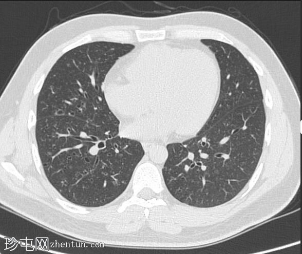
HRCT
CT
朗格汉斯细胞组织细胞增多症

Axial
non-contrast
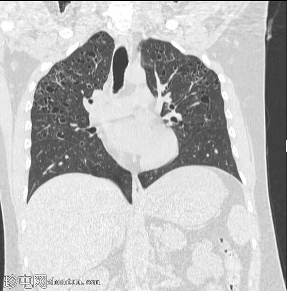
朗格汉斯细胞组织细胞增多症

Coronal
non-contrast
双侧多个形状怪异的囊肿,主要位于双上肺叶,不累及肺基部,伴有微妙的毛玻璃样混浊和一些小叶中心结节。
心脏大小正常。
大的血管并不引人注目。
没有肺部肿块的证据。
无心包或胸腔积液。
无气胸。
纵隔或腋窝无肿大淋巴结。
案例讨论
该患者有大量吸烟史,表现出朗格汉斯细胞组织细胞增多症的典型表现。
肺朗格汉斯细胞组织细胞增多症是一种上/中区主要疾病,不累及肋膈角。
参考
Zaveri J, La Q, Yarmish G, Neuman J. More Than Just Langerhans Cell Histiocytosis: A Radiologic Review of Histiocytic Disorders. Radiographics. 2014;34(7):2008-24. doi:10.1148/rg.347130132 - Pubmed
Meyer J & de Camargo B. The Role of Radiology in the Diagnosis and Follow-Up of Langerhans Cell Histiocytosis. Hematol Oncol Clin North Am. 1998;12(2):307-26. doi:10.1016/s0889-8588(05)70512-8
Sundar K, Gosselin M, Chung H, Cahill B. Pulmonary Langerhans Cell Histiocytosis. Chest. 2003;123(5):1673-83. doi:10.1378/chest.123.5.1673 - Pubmed |