fetus或foetus(/fiːtəs/;复数胎儿,feti,胎儿或foeti)是从胚胎发育的动物的未出生的后代。[1]胚胎发育后,发生胎儿发育阶段。在人类产前发育中,胎儿发育从受精后第9周(或第11周孕龄)开始,一直持续到出生。[2]产前发育是一个连续体,没有明确的定义特征区分胚胎和胎儿。然而,胎儿的特征在于所有主要身体器官的存在,尽管它们尚未完全发育和功能,并且一些尚未位于其最终解剖位置。
目录
1 词源
2 发展
2.1 9周至16周(2¼至4个月)
2.2 17周至25周(4¼至6¼个月)
2.3 26周至38(6½至9½个月)
2.4 增长的变化
3 生存能力
4 循环系统
4.1 出生前
4.2 产后发育
5 免疫系统
6 发展问题
7 胎儿疼痛
8 法律和社会问题
9 其他动物
10 参考资料
词源
fetus(复数胎儿)一词与拉丁语fētus(“后代”,“带来”,“孵化年轻”)[3] [4] [5]和希腊语“φυτώ”有关。英国,爱尔兰和英联邦的拼写是胎儿,至少从1594年开始使用。[6]带有-oe-的拼写出现在拉丁语晚期,其中元音之间的区别-oe-和-e-已经丢失。除了使用胎儿的医学文献外,这种拼写在大多数英联邦国家中最常见。在加拿大和美国使用更经典的拼写胎儿。此外,胎儿现在是医学期刊在全世界的标准英语拼写。[7]拼写faetus也被用于历史。[8]
发展
进一步的信息:产前发育
第9周至第16周(2至4个月)
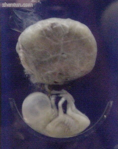
附着于胎盘的胎儿,胎龄3个月。
在人类中,胎儿阶段开始于第9周开始,[9]受精年龄或第11周孕龄。在胎儿阶段开始时,胎儿通常距离冠臀部约30毫米(1.2英寸),重约8克。[9]头部几乎占胎儿体重的一半。[10]胎儿的呼吸样运动对于刺激肺部发育是必要的,而不是用于获得氧气。[11]心脏,手,脚,大脑和其他器官都存在,但仅处于发育的开始阶段且操作极少。[12] [13]在第9周,胎儿的生殖器开始形成,胎盘完全发挥功能。[14]
在发育的这个阶段,随着肌肉,大脑和通路开始发展,不受控制的运动和抽搐发生。[15]
17至25周(4¼至6¼个月)
第一次怀孕的妇女(未经产)通常会在约21周时感觉到胎儿的运动,而之前分娩的妇女通常会感到运动20周。[16]到第五个月末,胎儿长约20厘米(8英寸)。
第26周至第38周(6½至9½个月)

艺术家在胎龄40周时描绘胎儿,从头到脚约51厘米(20英寸)。[17]
体脂肪量迅速增加。肺尚未完全成熟。形成了介导感官输入的丘脑大脑连接。骨骼完全发育,但仍然柔软和柔韧。铁,钙和磷变得更丰富。指甲到达指尖的尽头。胎毛或细毛开始消失,直到它消失,除了上臂和肩膀。两性都有小乳房芽。头发变得粗糙和浓密。出生即将来临,并在受精后第38周左右发生。胎儿在​​第36周和第40周之间被认为是足月,当它在子宫外的生命中充分发育时。[18] [19]出生时,它的长度可能是48到53厘米(19到21英寸)。出生时对运动的控制是有限的,有目的的自愿运动一直发展到青春期。[20] [21]
增长变化
更多信息:出生体重和环境毒物和胎儿发育
人类胎儿的生长存在很大差异。当胎儿体重小于预期时,该病症被称为胎儿宫内生长受限(IUGR),也称为胎儿生长受限(FGR);影响胎儿生长的因素可能是母体,胎盘或胎儿。[22]
母体因素包括孕妇体重,体重指数,营养状况,情绪压力,毒素暴露(包括烟草,酒精,海洛因和其他可能以其他方式伤害胎儿的药物)和子宫血流量。
胎盘因素包括大小,微观结构(密度和结构),脐带血流量,转运蛋白和结合蛋白,营养利用和营养产生。
胎儿因素包括胎儿基因组,营养物质产生和激素输出。此外,女性胎儿在足月时的体重往往低于男性。[22]
胎儿生长通常分类如下:小于胎龄(SGA),适合胎龄(AGA),大于胎龄(LGA)。[23] SGA可导致低出生体重,但早产也可导致低出生体重。低出生体重会增加围产期死亡率(出生后不久死亡),窒息,体温过低,红细胞增多症,低钙血症,免疫功能障碍,神经系统异常和其他长期健康问题的风险。 SGA可能与生长延迟有关,或者可能与绝对发育迟缓有关。
可行性
主要文章:胎儿活力

产前发育的阶段,显示生存能力和底部存活率为50%的点。几周和几个月由妊娠编号。
胎儿活力是指胎儿发育中胎儿可能在子宫外存活的点。生存率的下限约为孕龄5-3 / 4个月,通常较晚。[24]
胎儿自然变得可行的发育,年龄或体重没有明显限制。[25]根据2003 - 05年的数据,妊娠23周(5-3 / 4个月)出生的婴儿的存活率为20-35%; 24-25周(6 - 6-1 / 4个月)50-70%;并且在26-27周(6-1 / 2 - 6-3 / 4个月)及以上的时候> 90%。[26]对于体重小于1.1磅(0.50千克)的婴儿来说,很少能存活。[25]
当这样的早产儿出生时,死亡的主要原因是呼吸系统和中枢神经系统没有完全分化。如果给予专家的产后护理,一些体重低于1.1磅(0.50千克)的早产儿可能存活,并被称为极低出生体重或未成熟婴儿。[25]
早产是婴儿死亡的最常见原因,导致近30%的新生儿死亡。[26]在所有分娩的发生率为5%至18%时,[27]它也比早产更常见,其中妊娠占3%至12%。[28]
循环系统
主要文章:胎儿循环
出生前
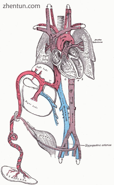
人胎儿循环系统的图。
循环系统的心脏和血管在胚胎发育期间相对较早地形成,但在生长的胎儿中继续生长和发展复杂。功能性循环系统是生物学必需品,因为哺乳动物组织在没有活跃血液供应的情况下不能生长超过几个细胞层厚度。产前血液循环不同于产后循环,主要是因为肺部未使用。胎儿通过胎盘和脐带从母体获得氧气和营养。[29]
来自胎盘的血液通过脐静脉运送到胎儿。其中约一半进入胎儿静脉导管并被带到下腔静脉,而另一半进入肝脏下缘的肝脏。供给右肝叶的脐静脉分支首先与门静脉相连。然后血液移动到心脏的右心房。在胎儿中,右心房和左心房(卵圆孔)之间有一个开口,大部分血液从右心腔流入左心房,从而绕过肺循环。大部分血流进入左心室,从那里通过主动脉泵入体内。一些血液从主动脉通过髂内动脉移动到脐动脉,并重新进入胎盘,胎儿的二氧化碳和其他废物被吸收进入妇女的血液循环。[29]
来自右心房的一些血液不进入左心房,而是进入右心室并被泵入肺动脉。在胎儿中,肺动脉和主动脉之间存在特殊的联系,称为动脉导管,它将大部分血液从肺部引导出来(此时胎儿悬浮在肺部,此时不用于呼吸)羊水)。[29]

3英寸(76毫米)胎儿的3D超声(约3-1 / 2个月胎龄)

胎儿在4-1 / 4个月

胎儿在​​5个月
产后发育
主要文章:适应宫外生活
出生后第一次呼吸,系统突然改变。肺阻力急剧减少,促使更多的血液从心脏的右心房和心室进入肺动脉,更少流过卵圆孔进入左心房。来自肺部的血液通过肺静脉进入左心房,产生压力增加,推动隔膜对着隔膜间隙,关闭卵圆孔并完成新生儿的循环系统分离到标准的左右两侧。此后,卵圆孔被称为卵圆窝。
动脉导管通常在出生后一两天内闭合,留下动脉韧带,而脐静脉和静脉导管通常在出生后2至5天内闭合,分别留下肝脏韧带和静脉韧带。
免疫系统
胎盘起到抵抗微生物传播的母胎屏障的作用。当这不足时,可能发生传染病的母婴传播。
母体IgG抗体穿过胎盘,使胎儿被动免疫,抵抗母亲有抗体的那些疾病。这种抗体在人类中的转移早在第五个月(孕龄)就开始,并且肯定在第六个月开始。[30]
发展问题
进一步的信息:环境毒物和胎儿发育和出生缺陷
发育中的胎儿极易受其生长和新陈代谢异常的影响,增加了出生缺陷的风险。一个值得关注的领域是怀孕期间的生活方式选择。[31]饮食在发育的早期阶段尤为重要。研究表明,用叶酸补充人的饮食可以降低脊柱裂和其他神经管缺陷的风险。另一个饮食问题是早餐是否被吃掉。不吃早餐可能会导致母体血液中的营养素长时间低于正常营养,导致早产或出生缺陷的风险增加。
酒精摄入可能会增加胎儿酒精综合症的发病风险,这是导致某些婴儿智力残疾的一种疾病。[32]怀孕期间吸烟也可能导致流产和低出生体重(2500克,5.5磅)。低出生体重是医疗服务提供者关注的问题,因为这些婴儿倾向于称为“过早体重”,具有较高的继发性医疗问题风险。
众所周知,X射线可能对胎儿的发育产生不利影响,需要权衡其风险。[33] [34]
一些研究表明,胎儿超声(包括多普勒,3D / 4D超声和2D超声)可能对出生体重和神经发育有一定影响。一个特别令人关注的问题是多年来胎儿超声波的广泛使用与自闭症病例数量的大量增加之间可能存在联系。[35]
先天性疾病是在出生前获得的。具有某些先天性心脏缺陷的婴儿只有在导管保持打开的情况下才能存活:在这种情况下,可以通过施用前列腺素来延迟导管的闭合,以允许有足够的时间来手术矫正异常。相反,在动脉导管未闭的情况下,导管未正确闭合,抑制前列腺素合成的药物可用于促使其闭合,从而可以避免手术。
其他心脏出生缺陷包括室间隔缺损,肺动脉闭锁和法洛四联症。
胎儿疼痛
主要文章:胎儿疼痛
胎儿疼痛[36],其存在及其影响在政治和学术上都有争议。根据2005年发表的一篇综述的结论,“关于胎儿疼痛能力的证据是有限的,但表明胎儿在妊娠晚期之前不太可能感觉疼痛。”[37] [38]然而,发育神经生物学家认为建立丘脑皮质连接(约6-1 / 2个月)是胎儿对疼痛感知的重要事件。[39]然而,疼痛的感知涉及感觉,情绪和认知因素,并且当经历疼痛时“不可能知道”,即使已知丘脑皮质连接已确立。[39]一些作者[40]认为妊娠后半期可能出现胎儿疼痛:“现有的科学证据表明胎儿疼痛感知可能在妊娠晚期之前发生,甚至可能,”IJP期刊中的KJS Anand写道。 [41]
胎儿是否有能力感受到疼痛和痛苦是堕胎辩论的一部分。[42] [43] 例如,在美国,支持生命的倡导者提出了立法,要求堕胎提供者告知孕妇,他们的胎儿在手术过程中可能会感到疼痛,并且需要每个人接受或拒绝胎儿的麻醉。[44]
法律和社会问题
在大多数国家,人工怀孕的流产是合法的和/或容忍的,尽管妊娠时间限制通常会禁止晚期堕胎。[45]
其他动物
进一步的信息:哺乳动物的进化

出生前大象发育的十四个阶段
胎儿是胎生生物产前发育的一个阶段。这个阶段介于胚胎发育和出生之间[1]。许多脊椎动物都有胎儿阶段,从大多数哺乳动物到许多鱼类。此外,一些无脊椎动物生活在幼年,包括一些种类的onychophora [46]和许多节肢动物。胎儿阶段趋同进化的普遍性表明它相对容易发展。它可能源于蛋的释放延迟,在产蛋之前卵在母体内孵化。随着时间的推移,蛋壳的坚固性可以降低,直到它变得只是一个囊。
大多数哺乳动物的胎儿与母亲的胎儿类似。[47]然而,与人类相比,携带垃圾的动物对胎儿周围区域的解剖结构是不同的:携带垃圾的动物的每个胎儿都被胎盘组织包围,并且沿着两个长子宫中的一个而不是单个子宫中的一个存在于子宫中。一个人类的女性。
出生时的发育在动物之间变化很大,甚至在哺乳动物中也不同。 Altricial物种在出生时相对无助,需要相当多的父母照顾和保护。相比之下,早熟的动物出生时眼睛睁得大大,有头发或向下,有大脑,并且可以立即移动并且有些能够逃避或保护自己抵御掠食者。除人类外,灵长类动物在出生时是早熟的。[48]
胎盘哺乳动物的妊娠持续时间从跳跃小鼠的18天到大象的23个月不等。[49]一般而言,较大陆地哺乳动物的胎儿需要较长的妊娠期。[49]

海豚的胎儿阶段
胎儿阶段的好处意味着年轻人在出生时更加发达。 因此,他们可能需要较少的父母照顾,并可能更好地自生自灭。 然而,携带胎儿会给母亲带来成本,母亲必须承担额外的食物以促进其后代的生长,并且其移动性和舒适性可能受到影响(特别是在胎儿期结束时)。
在某些情况下,胎儿阶段的存在可能允许有机体将其后代的出生时间定为有利的季节。[46]
另见:
Fetal position
Fetal rights
Fetoscopy
Neural development
Potential person
Pregnancy (mammals)
Superfetation
Women's rights
参考:
Ghosh, Shampa; Raghunath, Manchala; Sinha, Jitendra Kumar (2017), "Fetus", Encyclopedia of Animal Cognition and Behavior, Springer International Publishing, pp. 1–5, doi:10.1007/978-3-319-47829-6_62-1, ISBN 9783319478296
"First Trimester - American Pregnancy Association". americanpregnancy.org. 1 May 2012. Archived from the original on 23 April 2009.
O.E.D.2nd Ed.2005
Harper, Douglas. (2001). Online Etymology Dictionary Archived 2013-04-20 at the Wayback Machine. Retrieved 2007-01-20.
"Charlton T. Lewis, An Elementary Latin Dictionary, fētus". Archived from the original on 2017-01-04. Retrieved 2015-09-24.
"Foetus". Oxford English Dictionary. Archived from the original on February 17, 2019.
New Oxford Dictionary of English.
American Dictionary of the English Language, Noah Webster, 1828.
Klossner, N. Jayne, Introductory Maternity Nursing (2005): "The fetal stage is from the beginning of the 9th week after fertilization and continues until birth"
"Fetal development: MedlinePlus Medical Encyclopedia". www.nlm.nih.gov. Archived from the original on 2011-10-27.
Institute of Medicine of the National Academies, Preterm Birth: Causes, Consequences, and Prevention Archived 2011-06-07 at the Wayback Machine (2006), page 317. Retrieved 2008-03-12
The Columbia Encyclopedia Archived 2007-10-12 at the Wayback Machine (Sixth Edition). Retrieved 2007-03-05.
Greenfield, Marjorie. “Dr. Spock.com Archived 2007-01-22 at the Wayback Machine". Retrieved 2007-01-20.
Gavino, Dr Hazel (June 27, 2016). "9 Weeks Pregnant – Symptoms, Fetal Development, Tips". Archived from the original on 2016-08-16. Retrieved 2016-07-21.
Prechtl, Heinz. "Prenatal and Early Postnatal Development of Human Motor Behavior" in Handbook of brain and behaviour in human development, Kalverboer and Gramsbergen eds., pp. 415-418 (2001 Kluwer Academic Publishers): "The first movements to occur are sideward bendings of the head....At 9-10 weeks postmestrual age complex and generalized movements occur. These are the so-called general movements (Prechtl et al., 1979) and the startles. Both include the whole body, but the general movements are slower and have a complex sequence of involved body parts, while the startle is a quick, phasic movement of all limbs and trunk and neck."
Levene, Malcolm et al. Essentials of Neonatal Medicine (Blackwell 2000), p. 8. Retrieved 2007-03-04.
"Fetal development - 40 weeks". BabyCenter. 2015. Archived from the original on 29 August 2015. Retrieved 26 August 2015.
Your Pregnancy: 36 Weeks Archived 2007-06-01 at the Wayback Machine BabyCenter.com Retrieved June 1, 2007.
"full-term" defined by Memidex/WordNet.
Stanley, Fiona et al. "Cerebral Palsies: Epidemiology and Causal Pathways", page 48 (2000 Cambridge University Press): "Motor competence at birth is limited in the human neonate. The voluntary control of movement develops and matures during a prolonged period up to puberty...."
Becher, Julie-Claire. "Insights into Early Fetal Development". Archived from the original on 2013-06-01., Behind the Medical Headlines (Royal College of Physicians of Edinburgh and Royal College of Physicians and Surgeons of Glasgow October 2004)
Holden, Chris and MacDonald, Anita. Nutrition and Child Health (Elsevier 2000). Retrieved 2007-03-04.
Queenan, John. Management of High-Risk Pregnancy (Blackwell 1999). Retrieved 2007-03-04.
Halamek, Louis. "Prenatal Consultation at the Limits of Viability Archived 2009-06-08 at the Wayback Machine", NeoReviews, Vol.4 No.6 (2003): "most neonatologists would agree that survival of infants younger than approximately 22 to 23 weeks’ estimated gestational age [i.e. 20 to 21 weeks' estimated fertilization age] is universally dismal and that resuscitative efforts should not be undertaken when a neonate is born at this point in pregnancy."
Moore, Keith and Persaud, T. The Developing Human: Clinically Oriented Embryology, p. 103 (Saunders 2003).
March of Dimes - Neonatal Death Archived 2014-10-24 at the Wayback Machine, retrieved September 2, 2009.
World Health Organization (November 2014). "Preterm birth Fact sheet N°363". who.int. Archived from the original on 7 March 2015. Retrieved 6 March 2015.
Buck, Germaine M.; Platt, Robert W. (2011). Reproductive and perinatal epidemiology. Oxford: Oxford University Press. p. 163. ISBN 9780199857746. Archived from the original on 2016-08-15.
Whitaker, Kent. Comprehensive Perinatal and Pediatric Respiratory Care (Delmar 2001). Retrieved 2007-03-04.
Page 202 of Pillitteri, Adele (2009). Maternal and Child Health Nursing: Care of the Childbearing and Childrearing Family. Hagerstwon, MD: Lippincott Williams & Wilkins. ISBN 978-1-58255-999-5.
Dalby, JT (1978). "Environmental effects on prenatal development". Journal of Pediatric Psychology. 3 (3): 105–109. doi:10.1093/jpepsy/3.3.105.
Streissguth, Ann Pytkowicz (1997). Fetal alcohol syndrome: a guide for families and communities. Baltimore, MD: Paul H Brookes Pub. ISBN 978-1-55766-283-5.
O’Reilly, Deirdre. "Fetal development Archived 2011-10-27 at the Wayback Machine". MedlinePlus Medical Encyclopedia (2007-10-19). Retrieved 2018-08-26.
De Santis, M; Cesari, E; Nobili, E; Straface, G; Cavaliere, AF; Caruso, A (September 2007). "Radiation effects on development". Birth Defects Research. Part C, Embryo Today : Reviews. 81 (3): 177–82. doi:10.1002/bdrc.20099. PMID 17963274.
"Questions about Prenatal Ultrasound and the Alarming Increase in Autism - Midwifery Today". midwiferytoday.com. 1 December 2016. Archived from the original on 6 May 2012.
Lee, Susan J.; Ralston, Henry J. Peter; Drey, Eleanor A.; Partridge, John Colin; Rosen, Mark A. (2005-08-24). "Fetal Pain". JAMA. 294 (8): 947–54. doi:10.1001/jama.294.8.947. ISSN 0098-7484. PMID 16118385.
Lee, Susan; Ralston, HJ; Drey, EA; Partridge, JC; Rosen, MA (August 24–31, 2005). "Fetal Pain A Systematic Multidisciplinary Review of the Evidence". Journal of the American Medical Association. 294 (8): 947–54. doi:10.1001/jama.294.8.947. PMID 16118385. Archived from the original on 2008-01-10. Retrieved 2008-02-14. Two authors of the study published in JAMA did not report their abortion-related activities, which pro-life groups called a conflict of interest; the editor of JAMA responded that JAMA probably would have mentioned those activities if they had been disclosed, but still would have published the study. See Denise Grady, “Study Authors Didn't Report Abortion Ties” Archived 2009-04-25 at the Wayback Machine, New York Times (2005-08-26).
"Study: Fetus feels no pain until third trimester" Archived 2008-03-18 at the Wayback Machine MSNBC
Johnson, Martin and Everitt, Barry. Essential reproduction (Blackwell 2000): "The multidimensionality of pain perception, involving sensory, emotional, and cognitive factors may in itself be the basis of conscious, painful experience, but it will remain difficult to attribute this to a fetus at any particular developmental age." Retrieved 2007-02-21.
Glover V. The fetus may feel pain from 20 weeks. Conscience. 2004-2005 Winter;25(3):35-7
http://www.iasp-pain.org/AM/AMTe ... 90&SECTION=HOME Archived 2013-07-01 at the Wayback Machine
White, R. Frank. " [[New research has discovered that unborn babies can feel pain. "The neural pathways are present for pain to be experienced quite early by unborn babies,” explains Steven Calvin, M.D., perinatologist, chair of the Program in Human Rights Medicine, University of Minnesota, where he teaches obstetrics." [1]]http://www.asahq.org/Newsletters/2001/10_01/white.htm Are We Overlooking Fetal Pain and Suffering During Abortion?] Archived 2016-07-19 at the Wayback Machine", American Society of Anesthesiologists Newsletter (October 2001). Retrieved 2007-03-10.
David, Barry & and Goldberg, Barth. "Recovering Damages for Fetal Pain and Suffering Archived 2007-09-28 at the Wayback Machine", Illinois Bar Journal (December 2002). Retrieved 2007-03-10.
Weisman, Jonathan. "House to Consider Abortion Anesthesia Bill Archived 2008-10-28 at the Wayback Machine", Washington Post 2006-12-05. Retrieved 2007-02-06.
Anika Rahman, Laura Katzive and Stanley K. Henshaw. "A Global Review of Laws on Induced Abortion, 1985-1997 Archived 2016-03-03 at the Wayback Machine", International Family Planning Perspectives Volume 24, Number 2 (June 1998).
Campiglia, Sylvia S.; Walker, Muriel H. (1995). "Developing embryo and cyclic changes in the uterus ofPeripatus (Macroperipatus) acacioi (Onychophora, Peripatidae)". Journal of Morphology. 224 (2): 179–198. doi:10.1002/jmor.1052240207.
ZFIN, Pharyngula Period (24-48 h) Archived 2007-07-14 at the Wayback Machine. Modified from: Kimmel et al., 1995. Developmental Dynamics 203:253-310. Downloaded 5 March 2007.
Lewin, Roger. Human Evolution, page 78 (Blackwell 2004).
Sumich, James and Dudley, Gordon. Laboratory and Field Investigations in Marine Life, page 320 (Jones & Bartlett 2008). |