1.创建气腹
1.1启动气腹
气腹是最常用于腹腔镜暴露腹腔镜的方法。通过吹气扩张腹腔可提供足够的手术暴露,并允许腹腔镜手术所需的手术操作。建立气腹的最常见方式是吹入二氧化碳。
可以在两个基本过程中研究腹腔镜手术中与气腹相关的危险因素:
进入和暴露相关的危险因素和并发症气腹相关的改变和并发症
尽管已经开发出无气腹腔镜手术来克服气腹,尤其是孕妇(对胎儿)和老年患者的潜在不利影响,但二氧化碳(CO2)气腹仍然是最广泛使用的方法。
1.1.1进入腹腔和暴露
 
通常,二氧化碳以高速率(高达15L / min)吹入至12-16mmHg的压力极限。但是,根据年龄,体型和术中监测情况进行调整。作为压力指导方针:
婴儿:4-6 mmHg,吹气速度小于1 L / min
Child: 6–8 mmHg, insufflation rate around 1 L/min
Adult: 12–16 mmHg, insufflation rate less than 15 L/min
A variety of methods of primary peritoneal entry is available:
Noninsufflated entry method
Direct trocar and cannula
Open trocar and cannula
Pre-insufflated entry method with Veress needle
Conventional closed trocar and cannula
Shielded trocar and cannula
Radially expanding trocar and cannula
Visual entry method (with or without pre-insufflation)
Visual Veress needle
Visual disposable trocar and cannula
Visual reusable (EndoTIP) cannula
1.1.1.1 The Open Method
Open placement technique was first demonstrated in the United States by Hasson in 1975 and in Germany by Koenig in 1979. It is the safest method of initial port placement. Its use is not limited to initial port placement; any number of ports can be placed using this technique at any location in the abdomen. Some prefer to use this technique in selected cases such as slender, muscular patients, those with prior abdominal procedures or paediatric patients.
The initial port is usually placed at the umbilicus, as this is the thinnest part of the abdominal wall even in muscular or obese patients. If the patient has had a previous midline incision, the second most commonly accepted area to place the initial port is the left upper quadrant. However, placement off the midline in an obese individual can be dauntingly difficult or require an excessively large incision. Placing the incision within the umbilicus yields the most cosmetic scar. By placing the endoscope into the cannula but not through it, proper placement
can again be confirmed prior to insufflation. Hasson technique is particularly helpful in patients who have had multiple previous abdominal operations, in whom the risk of adhesions is increased. In some cases, adhesions may be so extensive as to require conversion to laparotomy.
1.1.1.2 The Veress Needle
Prior to blind placement of the Veress needle or trocars into the peritoneal cavity, the bladder and stomach are emptied, and the aorta palpated to decrease the chance of injury. The snap mechanism of the Veress needle is checked. A skin incision large enough to fit the ensuing trocar is made, and the subcutaneous tissues are bluntly dissected down to the anterior fascia. Generally, the spring mechanism will snap three times as the Veress needle penetrates the fascial layers and the peritoneum, while all resistance disappears once the peritoneal cavity is entered. The Veress needle should be freely mobile through 360°.
Testing
A number of tests have been devised to confirm placement in the peritoneal cavity. Injected saline flows freely and aspiration is freely accomplished returning neither blood nor enteric contents. The “slurp” test – A drop of saline placed in the closed Veress needle should flow freely once the needle is opened, especially if the abdominal wall is lifted.
The insufflation tubing may then be attached and started on low flow. Patient’s pressure reading should be low with free flow of gas and symmetric insufflation of the abdominal cavity. This serves as further confirmation of proper placement. A “visual Veress needle” is also on the market that further confirms proper placement using a thin endoscope that fits through the needle.
1.1.1.3 Trocar Placement Without Prior Pneumoperitoneum (Sharp/Bladeless)
Some surgeons advocate simple blind trocar placement with manual counter traction on the abdominal wall without prior pneumoperitoneum. However, it is better to use bladeless trocar under visual guidance of a laparoscope.
Regardless of the mode, once access is obtained, the first step is to inspect the peritoneal cavity to rule out iatrogenic injury.
1.2 Access and Exposure-Related Risk Factors
1.2.1 Improper Placement of the Veress Needle
Prevention
If one is not initially convinced of proper placement, due to failing one or more of the above-mentioned tests, one or two additional attempts at placement may be made. Then open placement technique should be undertaken.
1.2.2 Sudden Uncontrolled Entry into the Peritoneal Cavity
Prevention
Apply traction on the towel clips or directly retract the abdominal wall with one hand. Place the needle perpendicularly through the anterior abdominal wall under gentle controlled pressure, with the dominant hand resting on the abdominal wall, holding the needle not at the handle, but slightly closer to the tip.
1.2.3 High-Pressure Reading
If one is certain of proper placement but the pressure reading is too high, other causes for this include water in the tubing, closed valves, kinking of the tubing or inadequate relaxation of the patient.
Once the abdominal cavity is adequately insufflated, remove the needle, and place a sharp or dilating trocar with counter traction on the towel clips or by directly grasping and lifting the abdominal wall. After introducing the trocar, remove its inside part, keeping the outside cannula on place. Confirm the correct placement by the endoscope, and the insufflation tubing can be reconnected.
1.2.4 Poor Visualisation
Cause
Decreased light due to slightly bloodier nature of potential spaces or dirty scope.
Management
Increase intensity or gain of light; irrigate and suction the area. Frequently clean the lens with anti-fog solution and warm water as there are no proper peritoneal surfaces on which to wipe the scope.
1.2.5 Risks-Related Port Location
The most important factors in placing the ports are to space them in such a way that they do not interfere with each other and to trying to keep instruments in line with the camera. One rule of thumb is that the distance between the cannula and the operative site should be approximately half the length of the instrument. Ports are generally placed in a loosely configured semicircle around the operative site, although personal experience and specific patient factors may require a different number and configuration than routinely recommended. In the midline, the falciform ligament can get in the way superiorly, and the bladder can be injured inferiorly. More laterally, one risks injuring the epigastric vessels. Placing a port in the flanks risks damage to the colon. Excessive pressure at any site risks damage to all underlying organs and vessels.
Prevention
Transilluminate abdominal wall prior to trocar placement. The anatomy of the abdominal wall as well as underlying organs must be considered.
1.2.6 Uncontrolled Entry
Prevention
The skin incision should be one to two millimetres larger than the trocar. The trocar is generally placed with a twisting motion, applying pressure with the wrist, not the shoulder. The middle or index finger is extended along the trocar to prevent uncontrolled entry. The trocar is angled slightly towards the operative field, but not to a degree as for the trocar to slide along the outside of the peritoneum. Previously the “Z” placement technique was widely used to decrease leakage of pneumoperitoneum. Although this is less of a problem with newer trocars and grips, this technique may be useful in certain patients undergoing lengthy procedures.
1.2.7 Removal of Cannulas
Cannulas must be removed under direct visualisation, as a vessel may be tamponed with haemorrhage manifesting only after removal (Video 1.1). The operative field should be inspected under decreased pressure since bleeding may be tamponed by the pneumoperitoneum pressure and only become manifest once this is released.
Video 1.1: Trocar site bleeding 90 s
1.3 Access and Exposure-Related Complications
Complications of the Veress needle or blind placement techniques include vascular, gastrointestinal, urological and gynaecological trauma, as well as damage to solid organs. Although less frequent, these complications are not eliminated by using the open technique. If initial attempts to prevent or treat complications are unsuccessful, immediate cessation of the operation or conversion to laparotomy should be performed.
1.3.1 Preperitoneal Insufflation
Cause
Placement of the Veress needle at too great an angle or trocar sliding out into the preperitoneal space (Video 1.2).
Prevention
Place Veress needle perpendicularly and pass all tests, and place trocars securely and check placement prior to insufflation.
Management
Replace Veress needle or trocar, secure trocar with an additional fascial suture, convert to open procedure and allow time for resolution.
Video 1.2: Preperitoneal insufflation 54 s
1.3.2 Piercing the Greater Omentum with the Veress Needle
This can cause bleeding or produce interstitial emphysema of the greater omentum, pushing it against the anterior abdominal wall with insufflation.
Prevention
Make sure the abdominal wall is lifted during insertion of the Veress needle. Advance the tip less than one cm after the last audible snap of the Veress needle, rotate the needle and perform the safety tests.
Management
This complication is often recognised only after inserting the endoscope. Once recognised, withdraw the trocar to the level of the peritoneum then gently tap the abdominal wall from the outside to return the omentum to its original position.
Control of omental bleeding can usually be performed laparoscopically.
1.3.3 Puncture of a Hollow Organ with the Veress Needle
The “slurp” and rotation tests are generally not successful in this situation.
Prevention
Do not insert the Veress needle near laparotomy scars. Pay strict attention to all positioning tests.
Management
If you can bring the trocar back into the peritoneal cavity, an attempt to remove the carbon dioxide by aspiration through a fine needle may be made with laparoscopic repair of the injury. If unsuccessful, conversion to laparotomy is indicated.
1.4 Complications of Potential Space Exposure
Such areas include “the preperitoneal space” for hernia repair and urologic procedures; “the retroperitoneal space” for neurologic, vascular, orthopaedic or urologic procedures; and “the subfascial plane” in the leg for vascular procedures where no space actually exists. A variety of balloon dissectors is available. The principles of the procedure for entering the preperitoneal space may generally be applied to any area.
1.4.1 Haemorrhage
Prevention
Completely retract all strands of muscle laterally and cauterise any visible vessels.
1.4.2 Uneven Inflation
If the balloon inflates unevenly, or more dissection is needed unilaterally, manual pressure on the contralateral abdominal wall with further inflation of the balloon is sometimes of benefit.
1.4.3 Violation of the Peritoneal Cavity
Prevention
Direct the balloon dissector against the peritoneum when advancing it.
Management
Close the defect by suturing or clipping. Attempt placement on contralateral side or avoid defect and continue with procedures.
1.5 Complications of Trocar Placement
The visually controlled insertion of working ports for the operative instruments and retractors are important. More than 10 % of laparoscopic complications are associated with trocar insertion [6].
1.5.1 Abdominal Wall Haemorrhage
Cause
Laceration of abdominal wall vessel. The source of bleeding is usually the inferior epigastric artery or one of its branches.
Prevention
Transilluminate abdominal wall prior to trocar placement. Using the bladeless trocars minimises the risk by dilating the tissue while entering. Abdominal wall haemorrhage may be controlled with a variety of technique, including application of direct pressure with operating port, open or laparoscopic suture ligation, or apply pressure with a Foley catheter inserted into the peritoneal cavity.
Management
The trocar should be removed and the vessel cauterised externally or internally or ligated through an enlarged incision. The trocar is then replaced. Otherwise, the trocar may be removed with closure of the fascia and placement of the trocar elsewhere. Alternatively, a Foley balloon can be placed, inflated and retracted to tampon the bleeding. This method is time consuming, and if unsuccessful, one of the previously mentioned options must be undertaken. To understand the bleeding comes through which side, cantilever the trocar into each quadrant to find a position that causes the bleeding to stop. When the proper quadrant is found, pressure from the portion of the sheath within the abdomen tampons the bleeding vessel, thus stopping the bleeding. Then place a suture in such a manner that it traverses the entire border of the designated quadrant. Specialised
devices have been made that facilitate placement of a suture but are not always readily available. The needle should enter the abdomen on one side of the trocar and exit on the other side, thereby encircling the full thickness of the abdominal wall. This suture can be passed percutaneously either using a large curved # 1 absorbable suture as monitored endoscopically or using a straight Keith needle passed into the abdomen and then back out using laparoscopic grasping forceps. The suture, which encircles the abdominal wall, is tied over a gauze bolster to tampon the bleeding site.
1.5.2 Major Vascular Injury
Cause
Excessive pressure without adequate visualisation. Major vascular injury can occur when the sharp tip of the Veress needle or the trocar nicks or lacerates a mesenteric or retroperitoneal vessel. İt is rare when the open (Hasson cannula) technique is used.
Prevention
Controlled trocar placement under direct visualisation without undue pressure.
Management
If aspiration of the Veress needle reveals bloody fluids, remove the needle and puncture again the abdomen. Once access to the abdominal cavity has been achieved successfully, perform a full examination of the retroperitoneum to look for an expanding retroperitoneal haematoma. If there is a central or expanding retroperitoneal haematoma, laparotomy with retroperitoneal exploration is mandatory to access for and repair major vascular injury. Haematomas of the mesentery and those located laterally in the retroperitoneum are generally innocuous and may be just controlled by observation. If during closed insertion of the initial trocar there is a rush of blood through the trocar with associated hypotension, leave the trocar in place (to provide some tamponing of the haemorrhage and to assist in identifying the tract) and immediately perform laparotomy to repair what is likely to be an injury to the aorta, vena cava or iliac vessels. Minimal: pressure and thrombogens. Moderate or continued: if expertise is available, and the bleeding site clearly seen, then a maximum of two attempts may be made to control the bleeding by applying clips, ligatures or suturing. Heavy, continued or expertise not available: immediate conversion to laparotomy.
1.5.3 Bowel Injury
Prevention
Careful observation of the steps enumerated will minimise the chance of visceral injury. However, placement of the Veress needle is a blind manoeuvre, and even with extreme care, puncture of a hollow viscus is still possible. Once the peritoneal cavity has been entered, the trocar can be angled anteriorly to avoid potential danger to underlying organs.
Factors responsible for large vessel injury
Inexperienced or unskilled surgeon
Failure to sharpen the trocar
Failure to elevate or stabilise the abdominal wall
Perpendicular insertion of the needle or trocar
Lateral deviation of the needle or trocar
Inadequate pneumoperitoneum
Forceful thrust
Failure to note anatomical landmarks
Inadequate incision size
Management
If aspiration of the Veress needle returns yellowish or cloudy fluid, the needle is likely in the lumen of the bowel. Due to small calibre of the needle itself, this is usually a harmless situation. Simply remove the needle and puncture again the abdominal wall. After successful insertion of the laparoscope, examine the abdominal viscera closely for significant injury. If, however, the laparoscopic trocar itself lacerates the bowel, the injured area can sometimes be withdrawn through the incision and repaired extracorporeally or repaired intracorporeally depending on the experience of the surgeon. There are four possible courses of action: formal open laparotomy and bowel repair or resection; mini-laparotomy, using an incision just large enough to exteriorise the injured bowel segment for repair or resection and reanastomosis; laparoscopic resection of injured bowel and reanastomosis; and laparoscopic suture repair of the bowel injury. If possible, leave the trocar in place to assist in identifying the precise site of injury [7].
1.5.4 Bladder Injury
Prevention
Controlled trocar placement under direct visualisation, empty the bladder prior
to procedure.
Management
Damage to any organ is treated in a fashion similar to that for blood vessel injury. Two attempts if the expertise is available, otherwise, open. Drain the area and administer antibiotics as indicated.
1.6 Trauma Related with the Type of Port
The ports should be chosen with the specific procedure in mind, taking into account the surgeon’s preference, cost and what exact instrumentation will be needed.
There are many different trocar tips available, each with its own benefits and limitations. Pyramidal tips are reputed to cause more damage than conical tips; however, conical tips require excessive force to introduce. The knife blade tip theoretically causes less abdominal wall trauma; however, it cannot be introduced using the usual twisting method. Reusable trocars may become dull over time (Video 1.3).
Video 1.3: Injury risk related with the type of port 60 s
Many feel that shielded trocars are safer; however, they do not tend to be as easily introduced, leading to a greater amount of pressure being applied. The shield is supposed to pop out and lock around the blade once the trocar has entered the abdomen. However, if the skin incision is too small, the shield may be held back. Newer shielded trocars with a blade that retracts into the shield should eliminate this problem. Some newer cannulas minimise dangers associated with trocars. One is a disposable expandable sleeve, which is introduced using the Veress needle and then dilated to the necessary size using a blunt introducer. The other is a reusable threaded cannula, which is introduced through a small incision in the anterior fascia. Using rotational force, under direct vision, it bluntly dissects its way into abdomen. Another one is a disposable cannula with a bladeless trocar that is introduced under laparoscopic view by dilating the tissues. Both are reported to decrease trauma to the abdominal wall structures, as well as leaving smaller defects to close. No matter which product is used, careful controlled entry is the key.
1.7 Pneumoperitoneum-Associated Complications
1.7.1 Cardiopulmonary Trouble
Prevention
Keep the insufflation pressure and time to a minimum, proper patient selection.
Management
Evacuate the pneumoperitoneum. Cease the procedure if not too far advanced, convert to laparotomy if necessary, use adequate fluid resuscitation.
1.7.2 Gas Embolisation
Prevention
Use the lowest insufflation pressure compatible with adequate visualisation, reduce operating time, release pneumoperitoneum when not actively working.
Management
Evacuate pneumoperitoneum, left lateral decubitus position, 100 % oxygen, aspiration through central venous catheter, if catheter was previously placed [8].
1.7.3 Localised Collection of Carbon Dioxide
Manually deflate or aspirate prior to end of procedure.
1.7.4 Deep Vein Thrombosis
Prevention
Use antithrombotic pneumatic sleeves on lower extremities, low-dose heparin prophylaxis, keep pneumoperitoneum insufflation pressure and duration to a minimum.
1.7.5 Postoperative Shoulder or Subphrenic Pain
Cause
Alterations in the physiological environment of the peritoneal cavity may explain postoperative shoulder pain. Potential causes are the temperature of the gas used for the pneumoperitoneum as it leaves the storage cylinder (usually 20 °C), irritation of the diaphragm due to muscular distension as well as chemical reactions by gas on the peritoneum. Carbon dioxide is irritating to the peritoneum.
Prevention
Warming the gas to body temperature as it is insufflated and taking care to completely evacuate the peritoneal cavity may decrease pain. Other gases such as nitrous oxide have anaesthetic properties on the peritoneum; however, potential danger of combustion limits their widespread use.
Patients should be warned preoperatively that they may have shoulder pain (around 25 % of all patients) and that this will subside spontaneously within 2–3 days without analgesic treatment.
1.8 Pneumoperitoneum-Associated Physiologic Alterations
Cardiovascular/haemodynamic and pulmonary changes associated with the pneumoperitoneum represent a complex balance between the factors mentioned above. Carbon dioxide insufflation may also affect acid-base balance and may lead to further deterioration of existing intraperitoneal sepsis or inflammation. Factors Influencing Haemodynamic and Pulmonary Changes During laparoscopic Surgery
Mechanical effects of increased intra-abdominal pressure
Systemic effects of absorbed gas
Control of hypercarbia through augmentation of minute ventilation
Intravascular volume status
Body positioning (Trendelenburg and reverse-Trendelenburg positions)
Anaesthetic technique
Degree of surgical or pain stimulus
Cardiovascular comorbidity
1.9 Mechanical Effects of Increased Intra-abdominal Pressure
Insufflation of the abdominal cavity and elevation of intra-abdominal pressure have three predominant mechanical effects on cardiovascular functions:
Increased afterload
Increased venous resistance
Increased mean systemic pressure
Isolated elevation of intra-abdominal pressure produces compression of the splanchnic circulation, resulting in increased afterload and depression of cardiac function.
The increased abdominal pressure has an effect similar to positive end-expiratory pressure (PEEP) on haemodynamic variables. Thus, in animal studies, a decrease in cardiac output with concomitant increase of the central venous pressure and the peripheral systemic vascular resistance was observed after implementation of pneumoperitoneum [9]. Some other studies did not report on significant changes in cardiac output, while mean arterial pressure, systemic vascular resistance and central venous pressure were elevated [10].
In high-risk cardiac patients, the effect of CO2 pneumoperitoneum is more pronounced as compared to healthy individuals [11]. In addition, another study showed a decrease in heart rate and cardiac output without the compensatory mechanism of an elevated systemic venous resistance. The authors suggested that mixed venous oxygen saturation is the most sensitive parameter in monitoring cardiovascular function [12].
Pressure-related effects of pneumoperitoneum include decreased blood flow through the inferior vena cava. This in turn leads to reduced filling volume and pressure in the right and left atrium with consequent decrease in preload.
According to the Frank-Starling law, this effect can be compensated up to a point after which the cardiac output falls. Increases in central venous pressure due to higher intrathoracic pressure during mechanical ventilation and additional pneumoperitoneum falsely suggest sufficient volume status. Therefore, a decrease in cardiac output is the sequel of decreased preload, which is compensated by an increase in afterload from a rise in systemic venous resistance. The net effect is a stable or slightly decreased cardiac output and mean arterial pressure under normal conditions, i.e. adequate cardiac reserve and sufficient volume status. There is evidence that increasing intra-abdominal pressure decreases splanchnic blood flow, which adversely affects mucosal microcirculation.
Another study showed significant correlation between increasing intra-abdominal pressure and decreasing gastric mucosal pH measured by tonometry during CO2 pneumoperitoneum in a porcine model [13]. These results were interpreted as significant end-organ impairment, while at the same time significant differences in macrocirculatory parameters such as heart rate, cardiac output, pulmonary capillary wedge pressure and central venous pressure were not observed.
1.10 Direct Systemic Effects of Absorbed CO2
Transperitoneal absorption of CO2 is the main cause of hypercapnia when a CO2 pneumoperitoneum is established [14]. Hypercapnia has several effects on the cardiovascular system (Table 1.1).

Table 1.1 CO2 pneumoperitoneum-related effects on cardiovascular function
MAP mean arterial pressure, HR heart rate, SVR systemic vascular resistant, IP intraperitoneal pressure
Mild hypercapnia may lead to an increase in systemic venous resistance, thus increasing cardiac output and mean arterial pressure, while extensive hypercapnia causes depression of cardiac function [15]. After establishing a CO2 pneumoperitoneum, the systemic CO2 concentration rises due to the partial pressure difference between intraperitoneal CO2 and capillary blood pressure, leading to diffusion of CO2 into the blood. The resulting hypercapnia augments respiratory frequency and tidal volume in order to excrete the additional CO2. These compensatory mechanisms warrant an intact buffering system. In sick patients, however, additional CO2 might overwhelm this system and augment preexisting acidosis (e.g. in septic patients). Thus, a CO2 pneumoperitoneum in severe abdominal sepsis may be detrimental and should, therefore, be avoided [16]. This aspect may have significant implications considering reports on laparoscopic repair of hollow viscus perforations [17]. There are somewhat contradictory reports on changes in pO2 during laparoscopy. Effects of the pneumoperitoneum vary from decrease to no change to an increase in pO2. These discrepancies may be explained by the Trendelenburg position of the patient and different modes of ventilation between groups of patients. While patients who are breathing spontaneously show a lower pO2, patients on the
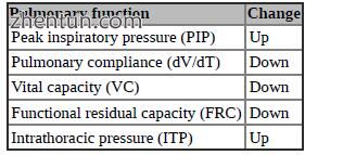
respirator have an elevated pO2 probably due to a higher FiO2. The changes in pulmonary function occurring during a CO2 pneumoperitoneum are summarised in Table 1.2 [18].

Table 1.2 CO2 pneumoperitoneum-related changes in pulmonary function
In comparison to an open operation, postoperative pulmonary function seems to be better after laparoscopy. Frazee and co-workers reported a better pulmonary function postoperatively in patients undergoing laparoscopic cholecystectomy as compared to patients undergoing open surgery [19].
1.11 Pneumoperitoneum and Abdominal Sepsis
Following the rapid acceptance of elective laparoscopic cholecystectomy, additional applications for minimal invasive surgery have been sought. Amongst these, laparoscopic closure of peptic ulcer perforation has been added to our operative armamentarium [17]. However, in conditions related to peritonitis, some experimental evidence has drawn attention to a theoretical risk concerning the CO2 pneumoperitoneum [20, 21].
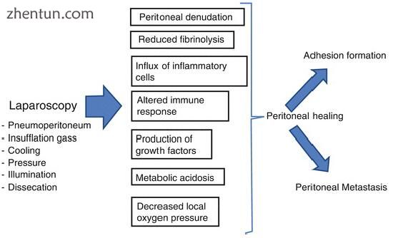
Laparoscopic surgical techniques require distension and elevation of the abdominal wall from the viscera to allow visualisation and manipulation. In the clinical situation, this is generally realised by intraperitoneal gas insufflation and maintenance of a continuous positive intra-abdominal pressure of approximately 8–12 mmHg. As observed in an experimental study, distension of the abdominal wall imposed by a pneumoperitoneum results in temporary stretching of the parietal mesothelial cells with concomitant flat bending of microvilli. Normal conformation of the mesothelium returns within 2 h after release of the pneumoperitoneum [16]. Increased intra-abdominal pressure due to the use of carbon dioxide insufflation apparently leads to the same ultrastructural changes observed after saline injection [22]. Thus, it may be concluded that it is the increased intra-abdominal pressure rather than a specific agent or gas that causes the described changes (Table 1.3).

Table 1.3 Laparoscopy-related peritoneal alterations
The parietal peritoneum physiologically functions as a barrier with controlled pathways to remove fluids, particles and cells from the peritoneal cavity. Abdominal secretions are drained by large terminal lymphatics that are located beneath the mesothelium of the peritoneal surface of the diaphragm. The absorbed fluid is then transported to the venous system by the thoracic duct. Increased intra-abdominal pressure has been shown to increase the resorption rate of intraperitoneal secretions [22, 23].
Furthermore, inflammatory stimuli are known to cause marked changes of the ultrastructural integrity of the mesothelial cell layer. Shrinking of mesothelial cells leads to disintegration and opening of the latticed intercellular network [22].
The combination of increased intra-abdominal pressure due to the CO2 pneumoperitoneum and of a gastric perforation with secondary inflammation results in premature deterioration of mesothelial integrity. The process of destruction includes numerical reduction as well as shrinking and coarsening of otherwise abundantly present microvilli. Furthermore, mesothelial cellular continuity is interrupted allowing the formation of stomata to the submesothelial cell layer. These changes to the ultrastructural anatomy of the mesothelial cell layer impair the barrier function of the parietal peritoneum giving way to uncontrolled resorption of abdominal secretions, which may induce bacteraemia, endotoxaemia and ultimately septic shock [16].
Based on these observations, experimental evidence demonstrating an aggravation of peritonitis and sepsis in conditions related to severe, long-lasting peritonitis is substantiated [20]. In contrast, another experimental setting in which the interval between bacterial inoculation and onset of pneumoperitoneum lasted only 60 min did not reveal any adverse effects [21].
Regarding both the available experimental and clinical evidence, a considerable risk for a pneumoperitoneum to aggravate peritonitis and to generate septic complications may be anticipated in conditions related to severe abdominal sepsis [24, 25]. Critical appraisal of laparoscopic surgery is warranted in conditions associated with severe, long-standing peritonitis.
参考:Complications in Laparoscopic Surgery A Guide to Prevention and Management |